Warning: mkdir(): Permission denied in /home/virtual/lib/view_data.php on line 81
Warning: fopen(upload/ip_log/ip_log_2024-04.txt): failed to open stream: No such file or directory in /home/virtual/lib/view_data.php on line 83
Warning: fwrite() expects parameter 1 to be resource, boolean given in /home/virtual/lib/view_data.php on line 84 A Study on the fine tegumental structures of the metacercaria and juvenile stages of Clonorchis sinensis
A Study on the fine tegumental structures of the metacercaria and juvenile stages of Clonorchis sinensis
Soon-Hyung Lee,Sung-Tae Hong and Byong-Seol Seo
Department of Parasitology and Institute of Endemic Diseases, College of Medicine, Seoul National University, Korea.
Abstract
This study was carried out to observe the chronological tegumental changes of juvenile C. sinensis using scanning and transmission electron microscopes (SEM and TEM). The subjected worms were the excysted metacercaria, and l day, 1 week, 2 week and 4 week old worms.
By observation with SEM, the tegument of excysted metacercaria showed many transverse wrinkling or shallow rugae, especially remarkable in anterior half of body, i.e., anterior to ventral sucker. Many spines were arrayed on the whole body surface, and double or triple pointed on anterior half and single pointed on posterior half. The observed sensory papillae were composed of 3 types. The ciliated knob-like papillae (type A in Fujino's classification) were abundant on anterior half, especially around oral sucker. A few plate like elevated papillae (type B) were found on middle part and non-ciliated round swellings (type C papillae) were observed around ventral sucker.
The tegumental surface of 1 day old worms showed deeper rugae, and the posterior body end was covered with cobble stone-like processes of distal cytoplasm. The spines protruded further and the spines on posterior half of body changed to scale-like ones.
The tegument of 1 week old worms became velvety and the spines grew further in length, but the density decreased.
The tegumental processes differentiated much finely in the 2 week and 4 week old specimens and the spines or sensory papillae decreased of their relative density.
The basic tegumental structures, such as distal cytoplasm containing various granules, vesicles and spines, basement membrane complex, muscular layers and tegumental cells were also monitored in 1 week old worms by TEM.
Figures
Figs. 1-5 Fig. 1. SEM, ventral view of whole excysted metacercaria (=EMC) of C. sinensis, showing two suckers, transverse wrinklings, sensory papillae and spines, ×790.
Fig. 2. SEM of C. sinensis EMC, posterior rim of oral sucker showing many ciliated sensory papillae (type A,), transverse rugae and spines, ×7,000.
Fig. 3. SEM of C. sinensis EMC, mid-portion between oral and ventral suckers showing numerous triple pointed spines, ×7,100.
Fig. 4. SEM of C. sinensis EMC, ventral surface of posterior body with numerous single pointed spines, and no tegumental rugae, ×7,100.
Fig. 5. SEM of C. sinensis EMC, oral sucker with many small or large sensory papillae, ×2,800.
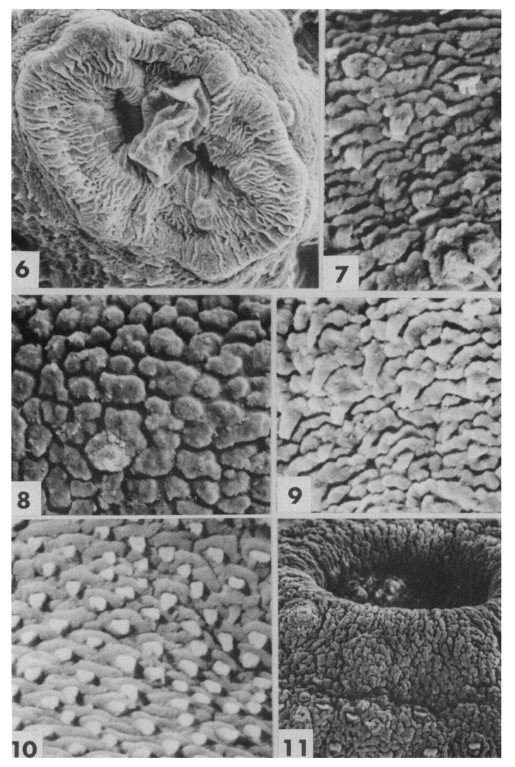
Figs. 6-11 Fig. 6. SEM of C. sinensis EMC, ventral sucker with several sensory papillae of type A and C (round swelling of sucker margin), ×2,530.
Fig. 7. SEM of 1 day old C. sinensis, dorsal surface showing more wrinkled tegument, double or triple pointed spines and cilliated sensory papillae, ×7,100.
Fig. 8. SEM of 1 day old C. sinensis, dorsal surface of posterior body covered by cobblestone-like tegumental processes, ×7,100.
Fig. 9. SEM of 1 day old C. sinensis, mid-dorsal surface with transverse rugae and spines, ×7,100.
Fig. 10. SEM of 1 day old C. sinensis, surface of posterior body with scale-like spines, ×7,100.
Fig. 11. SEM of 1 day old C. sinensis, oral sucker with many ciliated sensory papillae and the spines, ×5,100.
Figs. 12-16 Fig. 12. SEM of 1 week old C. sinensis, ventral surface posterior to oral sucker, showing paired ciliated sensory papillae, velvety tegumental processes and 3~4 pointed spines, ×7,100.
Fig. 13. SEM of 1 week old C. sinensis, velvety surface of posterior body with slender leaf-like spines, ×7,100.
Fig. 14. SEM of 1 week old C. sinensis, fine velvety bodysurface at the posterior end, ×7,100.
Fig. 15 & 16. TEM of 1 week old C. sinensis, showing the processes of tegumental syncytial layer, basement membrane, muscle layer and tegumental cell layer. A spine is sectioned in tegumental layer and numerous electron-dense granules are found, ×3,000.
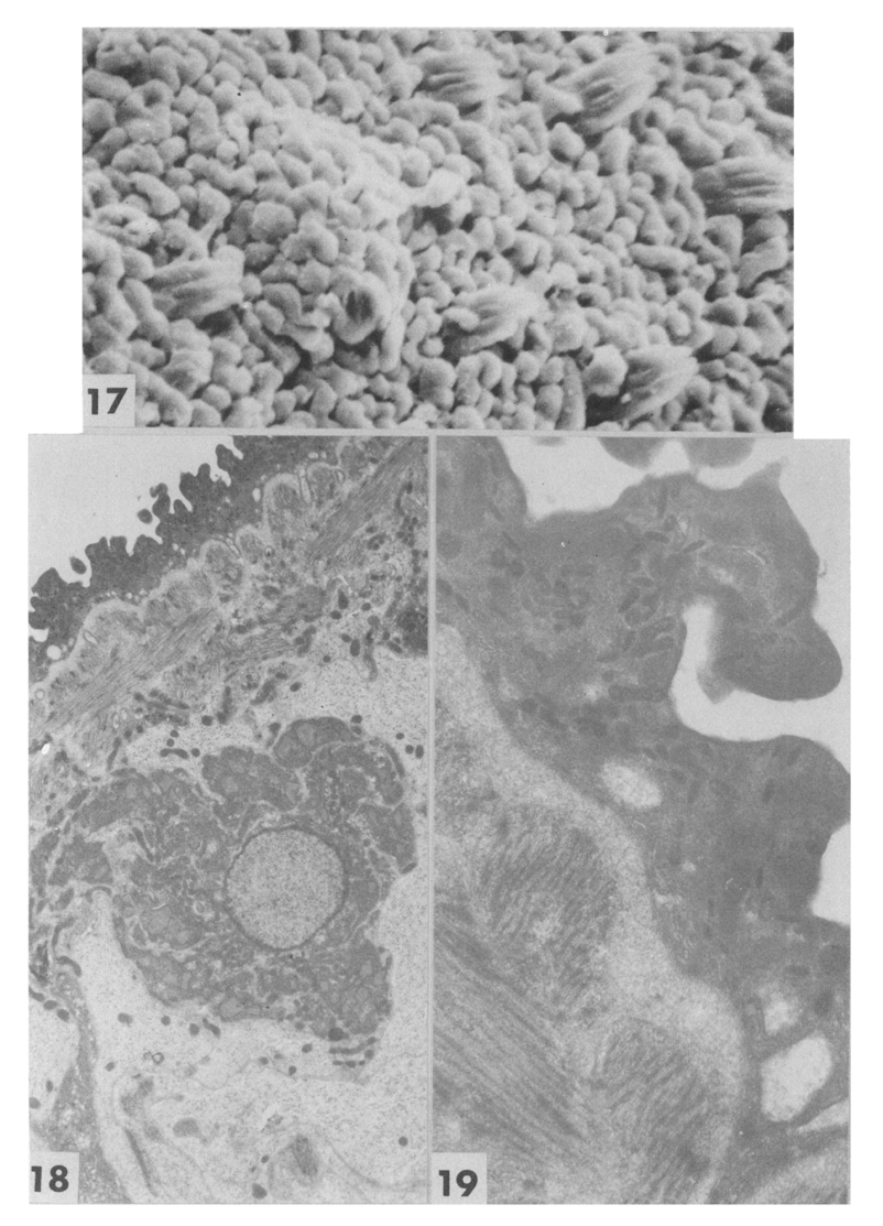
Figs. 17-19 Fig. 17. SEM of 2 week old C. sinensis, posterior to oral sucker, showing fine velvety tegumental processes with round tips and triple pointed spines, ×7,000.
Fig. 18. TEM of 2 week old C. sinensis, showing more differentiated tegumental syncytium, ×2,000.
Fig. 19. TEM of 2 week old C. sinensis, tegumental syncytium with many electron-dense granules and vacuoles, ×12,000.
Tables
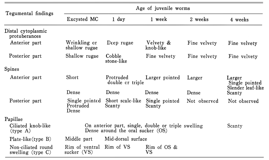
Table 1 Summary of SEM findings of C. sinensis tegument by the age of worms
References
1.
Fujino T, Ishii Y, Choi DW. The ultrastructural characterization of the tegument of Clonorchis sinensis (Cobbold, 1875) cercaria. Z Parasitenkd 1979;60(1):65–76.
2.
Fujino T, Ishii Y, Cho DW. Surface ultrastructure of the tegument of Clonorchis sinensis newly excysted juveniles and adult worms. J Parasitol 1979;65(4):579–590.
3.
Inatomi S, et al. Jpn J Parasit 1968;17:395–401.
4.
Jeong KH, Rim HJ, Kim CW. [A Study On The Fine Structure Of Clonorchis Sinensis, A Liver Fluke: 1. The Body Wall And The Nervous System]. Korean J Parasitol 1978;16(2):156–164.
5.
Kim CH. [Ultrastructure of the integument of adult Clonorchis sinensis]. Korean J Parasitol 1968;6(3):111–122.
6.
Kim SS, et al. Korea Univ Med J 1982;19:91–104.
7.
Saito T. Jpn J Parasit 1977;26:132–143.
8.
Seo BS, Lee SH, Cho SY, Chai JY, Hong ST, Han IS, Sohn JS, Cho BH, Ahn SR, Lee SK, Chung SC, Kang KS, Shim HS, Hwang IS. An Epidemiologic Study On Clonorchiasis And Metagonimiasis In Riverside Areas In Korea. Korean J Parasitol 1981;19(2):137–150.