Warning: mkdir(): Permission denied in /home/virtual/lib/view_data.php on line 81
Warning: fopen(upload/ip_log/ip_log_2024-04.txt): failed to open stream: No such file or directory in /home/virtual/lib/view_data.php on line 83
Warning: fwrite() expects parameter 1 to be resource, boolean given in /home/virtual/lib/view_data.php on line 84 A Study on the structure of Clonorchis sinensis, a liver fluke IV. Probable functions of the Laurer's canal
A Study on the structure of Clonorchis sinensis, a liver fluke IV. Probable functions of the Laurer's canal
Kye-Heon Jeong
Department of Biology, Soonchunhyang University, Onyang, Asan, Chungnam, Korea.
Abstract
A study on the function of the Laurer's canal of Clonorchis sinensis was conducted with help of the light microscope, the transmission electron microscope, and the scanning electron microscope. Some selected sexual organs concerning with the passages of the spermatozoa and the eggs were obseved in detail. The conclusion of this study is that the Laurer's canal may be the copulatory organ of the female reproductive system as Miyazaki et al. suggested in case of lung flukes.
Figures
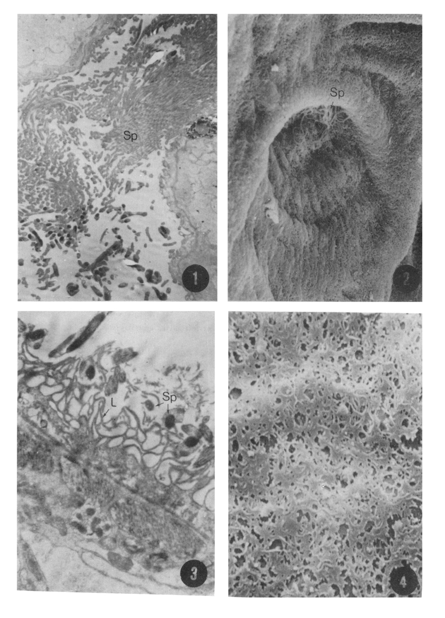
Figs. 1-4 Fig. 1. A transmission micrograph showing the mouth part of the seminal receptacle filled with numerous sperms. ×6,400
Fig. 2. A scanning electron micrograph showing the inner surface of the seminal receptacle with some sperms in the mouth part. ×1,500
Fig. 3. The epithelium of the seminal receptacle with well developed lamella. ×11,250
Fig. 4. An enlarged scanning electron micrograph of the seminal receptacle showing the lamellae in reticular arrangement. ×6,000
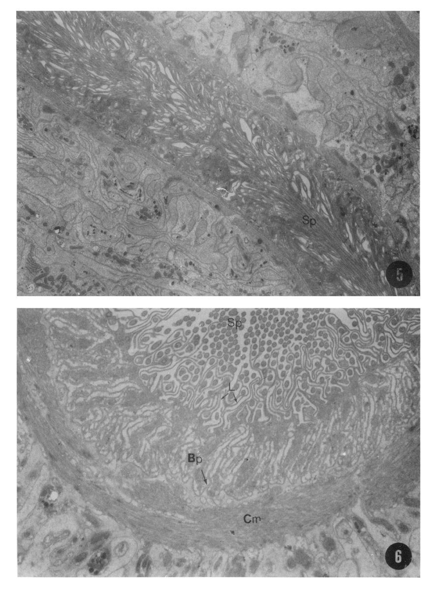
Figs. 5-6 Fig. 5. A transmission electron micrograph showing the sperm duct with well developed lamellae and a lot of sperms passing through the duct. ×3,000
Fig. 6. The cross sectioned view of the sperm duct, the basement plasma membrane deeply folded into the epithelium of the duct. ×8,000
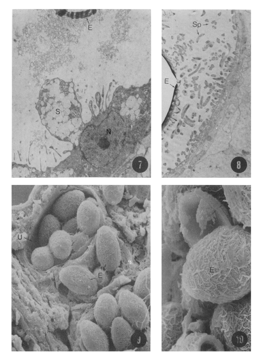
Figs. 7-10 Fig. 7. A transmission electron microscopic view showing the epithelium of the proximal uterus secreting egg shell materials. ×6,000
Fig. 8. A transmission electron micrograph showing the cervical uterus near the female opening with numerous sperms and eggs. ×1,500
Fig. 9. A scanning electron micrograph showing the numerous eggs pacted in the uterine tube. ×1,000
Fig. 10. A scanning electron micrograph showing a fractured egg. The thickness of the arrowed shell layer is about 1 µm. ×3,000
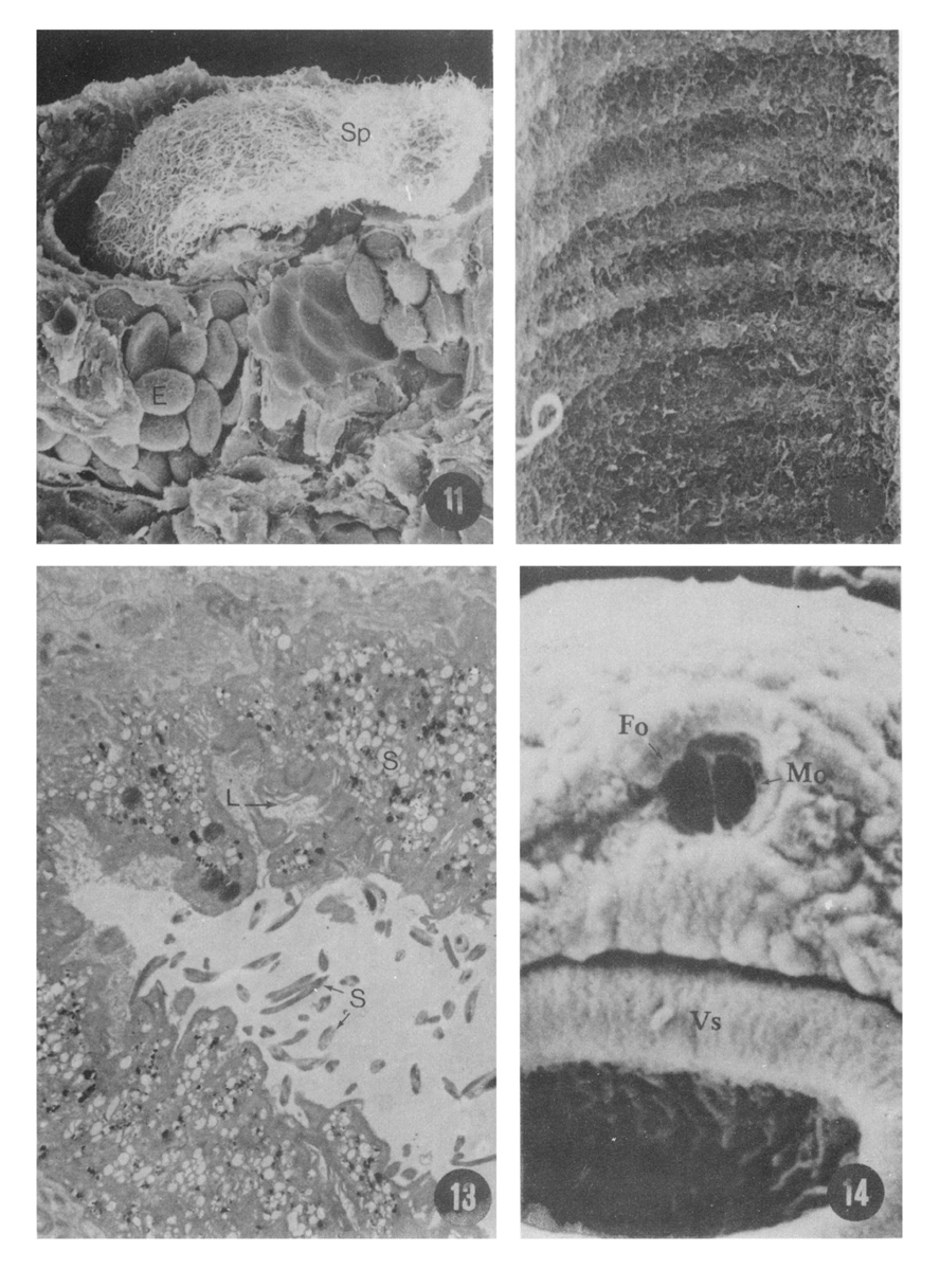
Figs. 11-14 Fig. 11. A scanning electron micrograph showing a lot of sperms passing through the vas deferens, and eggs in the uterus. ×450
Fig. 12. An enlarged scanning eletron micrograph showing the inner surface of the vas deferens. ×8,000
Fig. 13. A transmission electron microscopic view of the vas deferens showing the irregular inner surface with secretory materials from the prostate gland sperms in the lumen. ×11,600
Fig. 14. A scanning electron micrograph showing the genital atrium subdivided into male openings. ×1,040
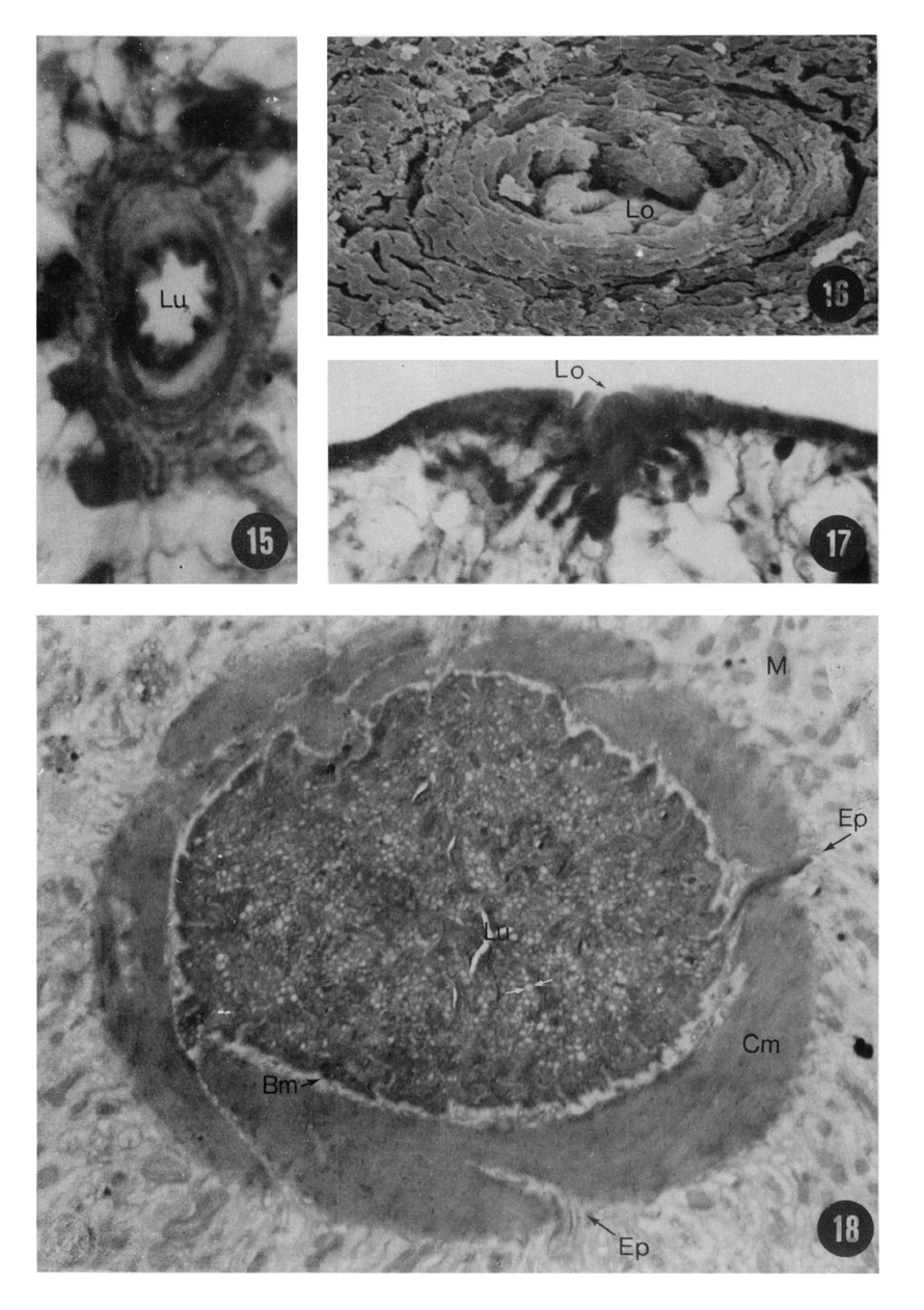
Figs. 15-18 Fig. 15. A light micrograph of the cross sectioned Laurer's canal showing the lumen. ×3,300
Fig. 16. A scanning electron micrograph showing the opening of the Laurer's canal on the dorsal side of the worm. ×3,000
Fig. 17. A light micrograph showing the opening of the Laurer's canal. ×2,300
Fig. 18. A transmission electron micrograph showing the cross sectioned view of the Laurer's canal. The well developed circular muscle bundles and the glandular epithelium are observed. ×8,600
Abbreviations
E
Egg
Bp
Basement plasma membrane
L
Lamella
Ep
Envagination of parenchyma
M
Mitochondria
Lo
Opening of the Laurer's canal
S
Secretory material
Lu
Lumen
U
Uterus
Mo
Male opening
Bm
Basement membrane
Sp
Sperm
Cm
Circular muscle
Vs
Ventral sucker
Fo
Female opening
References
1.
Anderson TF. Tr NewYork Acad Sci 1951;13:130–134.
2.
Faust EC, et al. Am J Hyg 1927;8:117.
3.
Fujino T, Ishii Y, Cho DW. Surface ultrastructure of the tegument of Clonorchis sinensis newly excysted juveniles and adult worms. J Parasitol 1979;65(4):579–590.
4.
Inatomi S. Okayama Igakkai zasshi 1962;74:71–75.
5.
Inatomi S, et al. Jpn J Parasit 1968;17:395–401.
6.
Jeong KH, Rim HJ, Yang HY, Kim WK, Kim CW. A Morphological Study On Spermatogenesis In The Liver Fluke, Clonorchis Sinensis. Korean J Parasitol 1976;14(2):123–132.
7.
Jeong KH, Rim HJ, Kim CW. [A Study On The Fine Structure Of Clonorchis Sinensis, A Liver Fluke: 1. The Body Wall And The Nervous System]. Korean J Parasitol 1978;16(2):156–164.
8.
Jeong KH, Rim HJ, Kim WK, Kim CW, Yang HY. [A Study On The Fine Structure Of Clonorchis Sinensis, A Liver Fluke: II. The Alimentary Tract And The Excretory System]. Korean J Parasitol 1980;18(1):81–91.
9.
Jeong KH, Rim HJ, Kim CW. [A Study On The Fine Structure Of Clonorchis Sinensis, A Liver Fluke: III.The Prostate Gland]. Korean J Parasitol 1980;18(1):93–97.
10.
Chu DS, et al. Korea Univ Med J 1982;19:71–77.
11.
Kim CH. [Ultrastructure of the integument of adult Clonorchis sinensis]. Korean J Parasitol 1968;6(3):111–122.
12.
Kang RS. Yonsei J Med Sci 1968;1:97–109.
13.
Kobayashi H. Dobutsugaku Zasshi 1910;22:1–4.
14.
Kobayashi H. Saikingaku Zasshi 1912;202:1–66.
15.
Kobayashi H. Centralbl Bakt Parasit Orrig 1912;75:299–318.
16.
Komiya Y, Kawana-Tajimi T. The development of the excretory system of Clonorchis sinensis in its definitive host. Jpn J Med Sci Biol 1953;6(6):571–575.
17.
Kusaura T. Med J Okayama Univ 1966;78:913–928.
18.
Paik KK, et al. Korean J Electr Micr 1969;1:35–42.
19.
Paik KK, et al. Korean J Electr Micr 1971;2:7–15.
20.
Takagi K. Tokushima J Exp Med 1962;9:60–66.
21.
Threadgold LT. Fasciola hepatica: the ultrastructure of the epithelium of the seminal vesicle, the ejaculatory duct and the cirrus. Parasitology 1975;71(3):437–443.
22.
Ujiie N. Taiwan Igakki Zasshi 1936;35:1,862–1,896.