Warning: mkdir(): Permission denied in /home/virtual/lib/view_data.php on line 81
Warning: fopen(upload/ip_log/ip_log_2024-04.txt): failed to open stream: No such file or directory in /home/virtual/lib/view_data.php on line 83
Warning: fwrite() expects parameter 1 to be resource, boolean given in /home/virtual/lib/view_data.php on line 84 In vitro effect of praziquantel on Paragonimus westermani by light and scanning electron microscopic observation
In vitro effect of praziquantel on Paragonimus westermani by light and scanning electron microscopic observation
Soon Hyung Lee,Ho Jin Park,Sung Jong Hong,Jong Yil Chai and Sung Tae Hong
Department of Parasitology and Institute of Endemic Diseases, College of Medicine, Seoul National University, Seoul 110, Korea.
Abstract
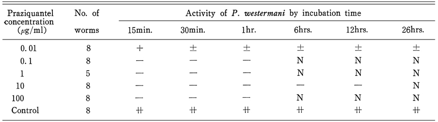
The effect of praziquantel on P. westermani exposed in vitro was observed by stereomicroscope, light microscope and scanning electron microscope. Following results were found. The worms incubated in 0.01 µg/ml praziquantel were moving after 26-hour incubation. However, all of them were immobilized immediately after incubation in solutions over 0.1 µg/ml concentration. All of the exposed worms showed severe vacuolization not only in tegument but in subtegument, intestine, ovary, testis, Mehlis' gland and excretory bladder. Vacuoles in tegument burst out to form craters. As incubation time went on, tegumental structure was disintegrated severely. The worms exposed to praziquantel were observed to be immobilized and be vacuolized of all tissues. Disintegration of reproductive organs suggests that praziquantel have suppressive effect on egg production when the flukes are not killed. The drug effects were found more related with incubation time than with drug concentration.
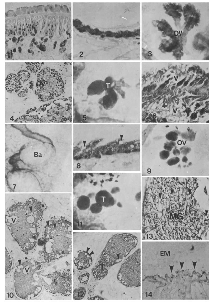
Fig. 1. Tegument of control worms, hematoxylin and eosin(GE) stained, ×200.
Fig. 2. Intestinal epithelium of a control worm, HE stained, ×200.
Fig. 3. A branched ovary(OV) of control worms with digited irregular margins, Semichon's acetocarmine(SA) stained, ×40.
Fig. 4. Ovary(OV) of a control worm. Note the compact germinal layer and medulla, HE stained, ×200.
Fig. 5. Testes(T) of control worms elongated with smooth marigin, SA stained, ×40.
Fig. 6. Antero-ventral tegument of a worm incubated in 10µg/ml praziquantel for 6 hours, with destruction of syncytium, muscle and subtegument by vacuoles(V), HE stained, ×400.
Fig. 7. Intestine of a worm incubated in 10µg/ml praziquantel for 6 hours showing constriction, and balloon (Ba) formation, and rough margin of wall, SA stained, ×40.
Fig. 8. Intestinal epithelium of a worm incubated in 10µg/ml praziquantel for 1 hour was thickened and vacuolized (arrow heads), HE stained, ×400.
Fig. 9. Ovary(OV) of a worm incubated in 10µg/ml praziquantel for 12 hours showing ball formation at distal part of every branch by constriction and disintegrated of proximal part, HE stained, ×40.
Fig. 10. Ovary of worm incubated in 0.1µg/ml praziquantel for 1 hour. Note large(V) and small vacuoles (arrow heads) in medulla, HE stained, ×100.
Fig. 11. Testes(T) of a worm incubated in 1µg/ml praziquantel for 1 hour. Note the single ball formed at the distal part of every testicular branch, HE stained, ×40.
Fig. 12. Testis(T) of a worm incubated in 0.1µg/ml praziquantel for 1 hour showed vaculoes(arrow heads) in periphery, HE stained, ×100.
Fig. 13. Mehlis' gland(MG) in 10µg/ml praziquantel for 6 hours was vacuolized(arrow heads), HE stained, ×200.
Fig. 14. Epithelium of excretory bladder showing shortened digitation(arrow heads) with eosinophilic excretion (EM) incubated in 0.1µg/ml praziquantel for 1 hour, HE stained, ×200.
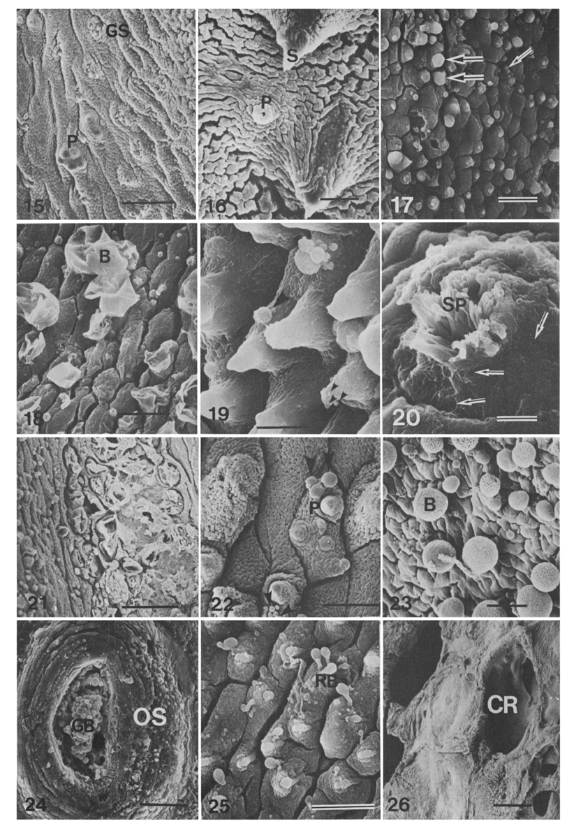
Figs. 15-26 Photograph of scanning electron microscopy(SEM) after gold coating.
Fig. 15. Ventral surface between oral and ventral suckers of a control worm showing velvety cytoplasmic processes, minute grouped spines(GS), and solitary or grouped papillae with a cilium(P), Bar=20µm, ×1,000.
Fig. 16. Postero-ventral tegument of a control worm. Note the cytoplasmic processes, single tipped spines(S) covered with cytoplasmic membrane and a solitary papilla(P) with a cilium, Mar=5µm, ×3,300.
Fig. 17. Beginning of bleb-formation(arrows) on the tegument between oral and ventral suckers of a worm incubated in 0.1µg/ml for 15 minutes, Bar=20µm, ×600.
Fig. 18. The ruptured blebs(B) and small ones on the anteroventral tegument of a worm incubated in 0.1µg/ml praziquantel for 1 hour, Bar=20µm, ×1,000.
Fig. 19. Minute blebs(arrow heads) on tips and grouped anes on base of spines in postero-ventral tegument of a worm incubated in 0.1µg/ml praziquantel for 1 hour, Bar=10µm, ×2,010.
Fig. 20. Genital atrium of a worm incubated in 0.1µg/ml praziquantel for 1 hour. Note the honeycimb-like craters(arrows) in tegument of dome and sperms(SP) in the genital opening, Bar=20µm, ×980.
Fig. 21. Disintegration of base of oral sucker by the ruptured blebs in a worm incubated in 1µg/ml praziquantel for 1 hour, Bar=20µm, ×990.
Fig. 22. Antero-ventral tegument of a wor incubated in 1µg/ml praziquantel for 1 hour. Note the blebs(arrow heads) around papillae(P) and the autolysis of swollen tegument, Bar=10µm, ×2,000.
Fig. 23. The wrinkled large blebs(B) on the postero-ventral tegument of a worm incubated in 10µg/ml praziquantel for 30 minutes, Bar=30µm, ×500.
Fig. 24. A worm incubated in 10µg/ml praziquantel for 1 hour, the inner and outer tegument of oral sucker(OS) was destroyed by numerous blebs and a grape-like blebs(GB) appeared in oral cavity, Bar=50µm, ×300.
Fig. 25. A few rubber bulb-like (club-shaped) blebs(RB) which had cylindrically extended tubular proximal part were observed on the ventrolateral tegument of a worm incubated in 10µg/ml praziquantel for 1 hour, Bar=20µm, ×1,010.
Fig. 26. The very large craters(CR) reaching to basement membrane layer and smooth surface disintegrated by the vaculoes formed from syncytium was observed in ventrolateral tegument of a worm incubated in 10µg/ml praziquantel for 6 hours, Bar=20µm, ×720.
Tables
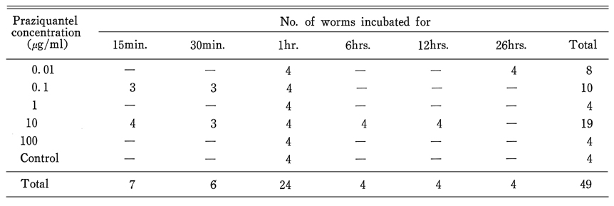
Table 1 Number of P. westermani prepared for light microscopy by prraziquantel concentration and incubation time
Table 2 Number of P. westermani prepared for scanning electron microscopy by praziquantel concentration and incubation time
Table 3 Chronological observations on the activity of P. westermani incubated in various concentrations of praziquantel solution
References
1.
Andrews P, Thomas H, Weber H. The in vitro uptake of 14C-praziquantel by cestodes, trematodes, and a nematode. J Parasitol 1980;66(6):920–925.
2.
Becker B, et al. Z Parasitenkd 1980;83:113–128.
3.
Chiu HS, et al. Korea Univ Med J 1982;19:617–630.
4.
Choi WY, Yoo JE. [Ultrastructure of the integument of adult Paragonimus westermani]. Korean J Parasitol 1985;23(1):111–122.
5.
Davis A, Biles JE, Ulrich AM. Initial experiences with praziquantel in the treatment of human infections due to Schistosoma haematobium. Bull World Health Organ 1979;57(5):773–779.
6.
Higo H, et al. Jpn J Parasit 1984;33:421–427.
7.
Hong ST, Lee SH, Ahn HS, Yun CK. A Case Of Niclofolan (Bilevon(R)) Intoxication. Korean J Parasitol 1982;20(1):49–52.
8.
Kim SS, et al. Korea Univ Med J 1982;19:91–105.
9.
Lee GJ. J Catholic Med College 1984;37:433–455.
10.
Lee SH. Seoul J Med 1985;26:41–51.
11.
Lee SH, Seo BS, Chai JY, Hong SJ. [Study on Metagonimus yokogawai(Katsurada, 1912) in Korea VII. Electron microscopic observation on the tegumental structure]. Korean J Parasitol 1984;22(1):1–10.
12.
Lee SM, et al. Chung-Ang J Med 1981;6:579–589.
13.
Mehlhorn H, Becker B, Andrews P, Thomas H, Frenkel JK. In vivo and in vitro experiments on the effects of praziquantel on Schistosoma mansoni. A light and electron microscopic study. Arzneimittelforschung 1981;31(3a):544–554.
14.
Mehlhorn H, Kojima S, Rim HJ, Ruenwongsa P, Andrews P, Thomas H, Bunnag B. Ultrastructural investigations on the effects of praziquantel on human trematodes from Asia: Clonorchis sinensis, Metagonimus yokogawai, Opisthorchis viverrini, Paragonimus westermani and Schistosoma japonicum. Arzneimittelforschung 1983;33(1):91–98.
15.
Rim HJ. Korea Univ Med 1975;12:425–457.
16.
Rim HJ, et al. Korea Univ Med 1980;17:113–128.
17.
Rim HJ, Kim MS, Ha JH, Chang DS. Expermental chemotherapeutic effects of niclofolan (Bayer 9015, Bilevon) on the animals infected with Paragonimus westermani or P. iloktsuenensis. Korean J Parasitol 1976;14(2):140–146.
18.
Rim HJ, Lyu KS, Lee JS, Joo KH. Clinical evaluation of the therapeutic efficacy of praziquantel (Embay 8440) against Clonorchis sinensis infection in man. Ann Trop Med Parasitol 1981;75(1):27–33.
19.
Seo BS, Cha IJ, Chai JY, Hong SJ, Lee SH. Studies on intestinal trematodes in Korea XIX. Light and scanning electron microscopy of Fibricola seoulensis collected from albino rats treated with praziquantel. Korean J Parasitol 1985;23(1):47–57.
20.
Sirisinha S, Puengtomwatanakul S, Sobhon P, Saitongdee P, Wongpayabal P, Mitranonde V, Radomyos P, Bunnag D, Harinasuta T. Alterations of the surface tegument of Opisthorchis viverrini exposed to praziquantel in vitro and in vivo. Southeast Asian J Trop Med Public Health 1984;15(1):95–103.