Warning: mkdir(): Permission denied in /home/virtual/lib/view_data.php on line 81
Warning: fopen(upload/ip_log/ip_log_2024-04.txt): failed to open stream: No such file or directory in /home/virtual/lib/view_data.php on line 83
Warning: fwrite() expects parameter 1 to be resource, boolean given in /home/virtual/lib/view_data.php on line 84 A study on the body fluid antigen of Clonorchis sinensis using immunogold labeling method
A study on the body fluid antigen of Clonorchis sinensis using immunogold labeling method
B D Chu,H J Rim and S J Kim*
Department of Parasitology and Institute for Tropical Endemic Diseases, College of Medicine, Korea University, Seoul 110-702, Korea.
*Department of Biology, Hallym University, Chunchon 200-702, Korea.
Abstract
In order to observe the antigenic localization in the tissues of the adult Clonorchis sinensis, immunogold labeling method was applied using serum immunoglobulins (IgG) of either worm-infected rabbits (group I) or antigen-immunized rabbits (group II) (by the body fluid obtained from the adult worms). The electron micrographs of the sectioned worm tissue antigens, embedded in Lowicryl HM 20 medium and stained with protein A-gold complex (particle size: 12 nm), were compared between the group I and group II. The gold particles were observed in the interstitial matrix of the worm parenchyma, the epithelial lamellae of the cecum, and the cecal lumen both in group I and II. But the particles were in general more concentrated in group II.
The gold particles were not observed on the basal lamina of the tegument or on vitelline glands in group I, while they were highly concentrated on those areas in group II.
There were also differences in the antigenicity of interstitial matrix(reacted with group I IgG) and head part(reacted with group II IgG) of the sperm cells in the seminal receptacle.
Conclusively, it is suggested that the substances comprising the basal lamina of the tegument or vitelline glands act as specific antigens reacting with antigen(body fluid) immunized rabbit IgG. On the other hand, the substances in the cecal lumen and cecal epithelial lamellae are thought to be the specific antigen that react with the worm-infected rabbit IgG.
Figures
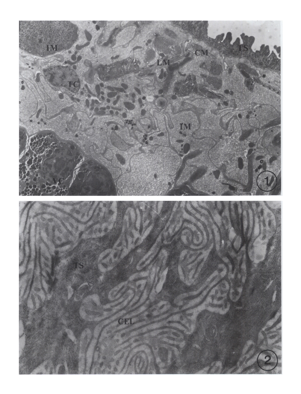
Figs. 1-2 Electron micrographs of sections of the worm tissue which were stained with protein A-gold complex. Gold particle size, 12 nm.
Fig. 1. The tegument of the worm, which was reacted with normal rabbit IgG, is composed of the tegumental syncytium(TS), basal layer(BL), circular(CM) and longitudinal(LM) muscle layers, interstitial matrix(IM) and tegumental cells(TC). A lot of multiple granules are accumulated in the tegumental syncytium of the worm. Gold particles are not found in the tegument or other partions of the tissue (× 5,600).
Fig. 2. Electron micrograph of a cecal section of a worm reacted with normal rabbit IgG. Gold particles are not found in the lumen or epithelial (CEL) of the cecum (× 30,000)
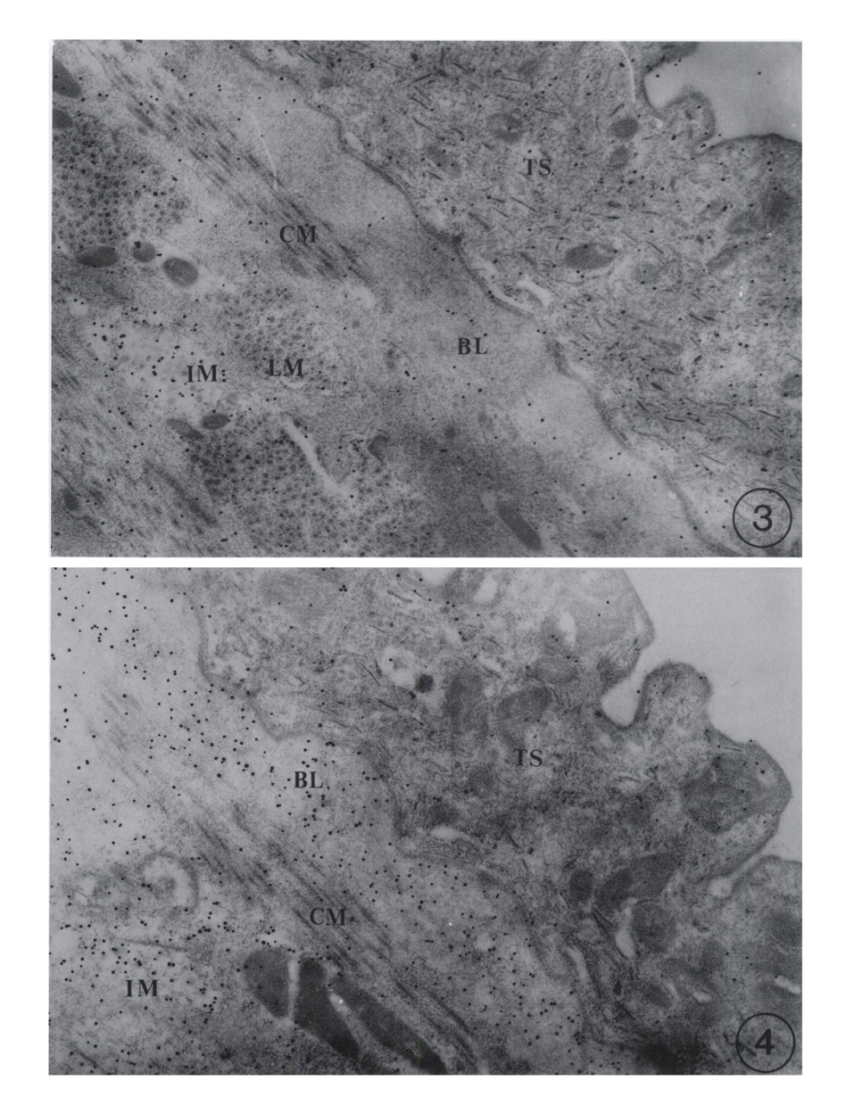
Figs. 3-4. Electron micrographs of sections of the worm tissue which were stained with protein A-gold complex. Gold particle size, 12 nm.
Fig. 3, 4. Gold particles are labeled in the interstitial matrix of the tegument of the worm which was reacted with infected rabbit IgG (Fig. 3). But in the tegument of the worm which was reacted with immunized rabbit IgG, the interstitial matrix and basal layer of the tegument are labeled (Fig. 4). (× 30,000 for Figs. 3 and 4)
Figs. 5-6 Electron micrographs of sections of the worm tissue which were stained with protein A-gold complex. Gold particle size, 12 nm.
Fig. 5, 6. Electron micrographs of a cecal section of the worms reacted with infected rabbit IgG (Fig. 5) or immunized rabbit IgG (Fig. 6). Gold particles are concentrated over the cecum epithelial lamellae(CEL) and lumen matrix of the cecum (× 30,000) for Figs. 5 and 6).
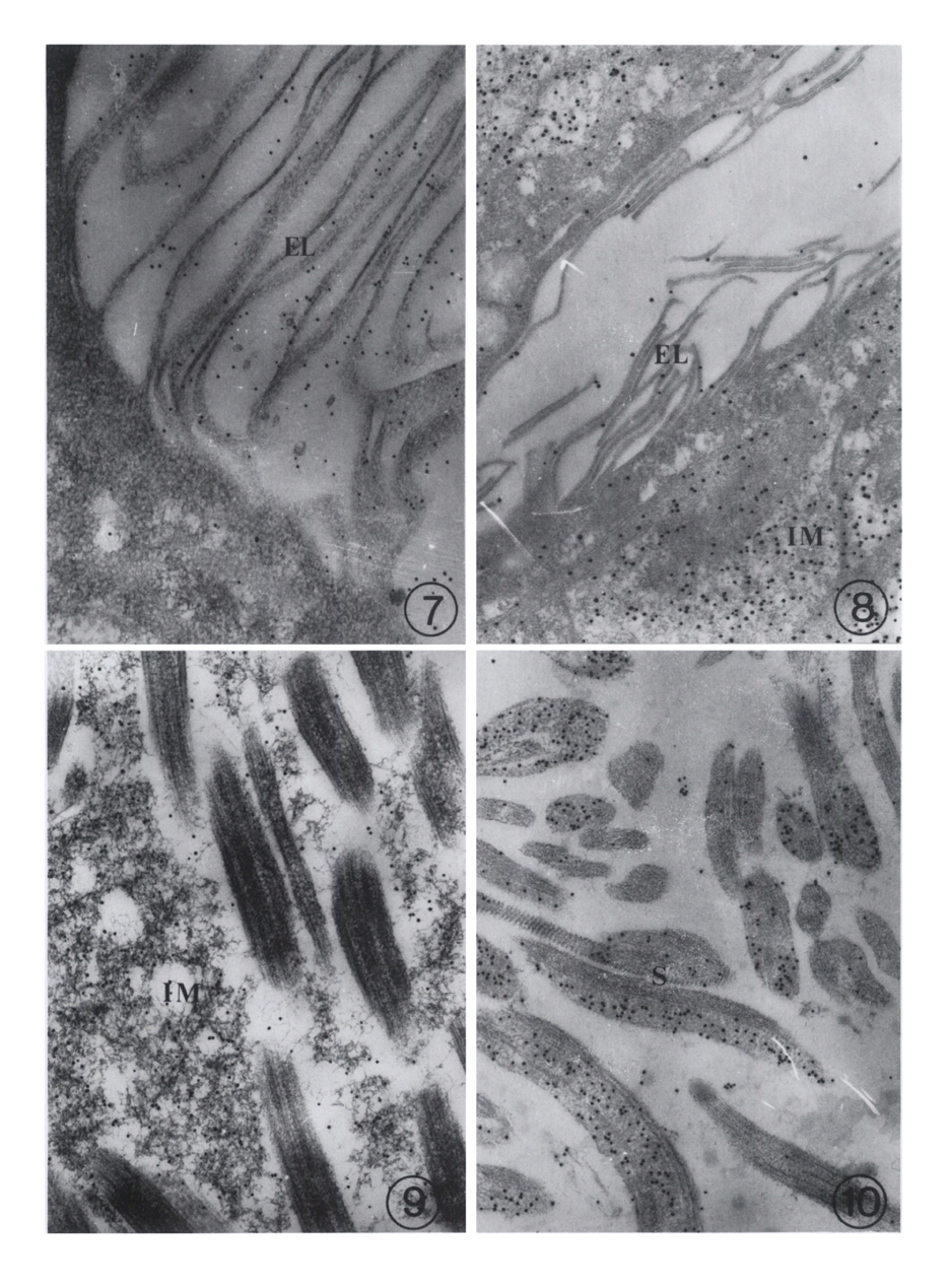
Figs. 7-10 Electron micrographs of sections of the worm tissue which were stained with protein A-gold complex. Gold particle size, 12 nm.
Fig. 7, 8. Electron micrographs of section of excretory cannal of the worm reacted with infected rabbit IgG (Fig. 7) or immunized rabbit IgG (Fig. 8). Gold particles are concentrated in epithelial lamellae(EL) of the excretory cannal and excretory bladder (Fig. 7), whereas the excretory cannal of the worm which was reacted with immunized rabbit IgG is not labeled in epithelial lamellae(EL) and in lumen (Fig. 8) (× 30,000 for Figs. 7 and 8).
Fig. 9, 10. Electron micrographs of section of receptaculum seminis of the worm reacted with infected rabbit IgG (Fig. 9) or immunized rabbit IgG. The receptaculum seminis which was reacted with infected IgG is labeled over the interstitial matrix (Fig. 9) but receptaculum seminis which was reacted with immunized IgG is labeled in the sperm head (Fig. 10) (× 30,000 for Figs. 9 and 10).
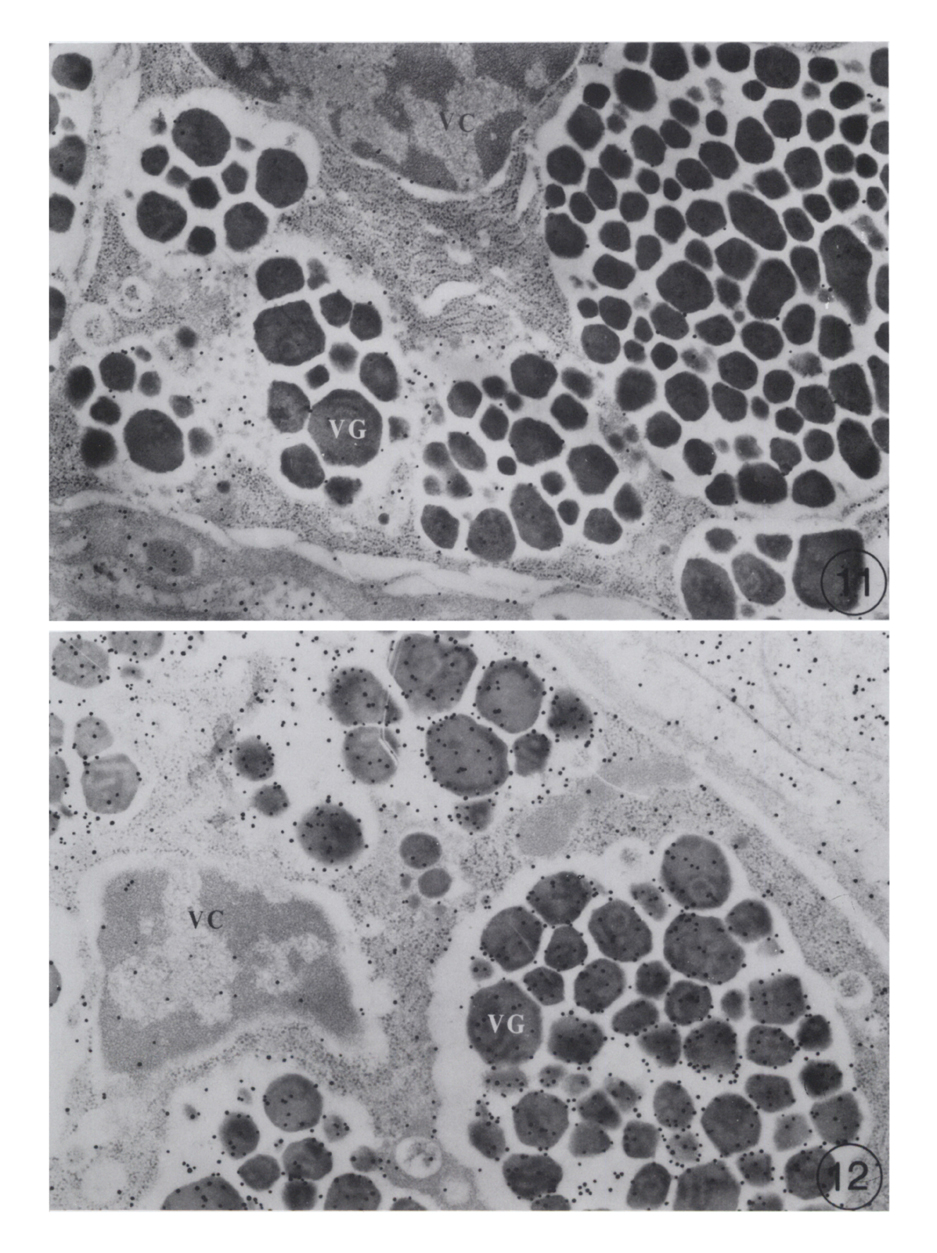
Figs. 11-12 Electron micrographs of sections of the worm tissue which were stained with protein A-gold complex. Gold particle size, 12 nm.
Fig. 11, 12. Electron micrographs of section of vitelline gland of the worm which was reacted with infected (Fig. 11) or immunized IgG. The vitelline glands are not labeled in their globules in the former (Fig. 11), but strongly labeled in the latter (Fig. 12). (× 30,000 for Figs. 11 and 12).
Tables
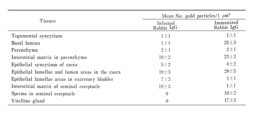
Table 1 Quantiative density of the labeled gold particles in the tissues of C. sinensis reacted with antibodies (IgG)* obtained from rabbits infected with C. sinensis or rabbits immunized with the body fluid of C. sinensis
References
1.
Bretana A, Avila JL, Arias-Flores M, Contreras M, Tapia FJ. Trypanosoma cruzi and American Leishmania spp: immunocytochemical localization of a laminin-like protein in the plasma membrane. Exp Parasitol 1986;61(2):168–175.
2.
Cho KM, et al. Yonsei Rep Trop Med 1974;5:45–56.
3.
Cho KM, et al. Yonsei Rep Trop Med 1976;7:26–39.
4.
Chu DS, et al. Korea Univ Med J 1982;19:71–80.
5.
Chung BJ, Joo CY, Choi DW. Seasonal variation of snail population of Parafossarulus manchouricus and larval trematode infection in river Kumho, Kyungpook province, Korea. Korean J Parasitol 1980;18(1):54–64.
6.
Jeong KH, Rim HJ, Kim CW. [A Study On The Fine Structure Of Clonorchis Sinensis, A Liver Fluke: 1. The Body Wall And The Nervous System]. Korean J Parasitol 1978;16(2):156–164.
7.
Jeong KH, Rim HJ, Kim WK, Kim CW, Yang HY. [A Study On The Fine Structure Of Clonorchis Sinensis, A Liver Fluke: II. The Alimentary Tract And The Excretory System]. Korean J Parasitol 1980;18(1):81–91.
8.
De Duve C, Beaufay H. A short history of tissue fractionation. J Cell Biol 1981;91(3 Pt 2):293s–299s.
9.
Erasmus DA. Schistosoma mansoni: development of the vitelline cell, its role in drug sequestration, and changes induced by Astiban. Exp Parasitol 1975;38(2):240–256.
10.
Erasmus DA, Davies TW. Schistosoma mansoni and S. haematobium: calcium metabolism of the vitelline cell. Exp Parasitol 1979;47(1):91–106.
11.
Erasmus DA, Popiel I. Schistosoma mansoni: drug induced changes in the cell population of the vitelline gland. Exp Parasitol 1980;50(2):171–187.
12.
Fujino T, Ishii Y, Cho DW. Surface ultrastructure of the tegument of Clonorchis sinensis newly excysted juveniles and adult worms. J Parasitol 1979;65(4):579–590.
13.
Fukuda K, et al. Jpn J Parasitol 1983;32(5):439–449.
14.
Gupta AN, Guraya SS, Sharma PN. Histochemical observations on the excretory system of digenetic trematodes. Acta Morphol Neerl Scand 1974;12(3):231–242.
15.
Hanna RE. Fasciola hepatica: an electron microscope autoradiographic study of protein synthesis and secretion by gut cells in tissue slices. Exp Parasitol 1975;38(2):167–180.
16.
Kim SJ, et al. Korean J Zool 1988;31:62–70.
17.
Kim SJ, Lee KO, Takamiya S, Capaldi RA. Mitochondrial myopathy involving ubiquinol-cytochrome c oxidoreductase (complex III) identified by immunoelectron microscopy. Biochim Biophys Acta 1987;894(2):270–276.
18.
von Lichtenberg F, Bawden MP, Shealey SH. Origin of circulating antigen from the schistosome gut. An immunofluorescent study. Am J Trop Med Hyg 1974;23(6):1088–1091.
19.
Madhavi R, et al. Rivista di Parasitol 1972;33:173–182.
20.
Madhavi R, et al. Rivista di Parasitol 1974;35:23–36.
21.
Palade G. Intracellular aspects of the process of protein synthesis. Science 1975;189(4200):347–358.
22.
Parshad VR, Guraya SS. Comparative histochemical observations on the excretory system of helminth parasites. Z Parasitenkd 1977;52(1):81–89.
23.
Roth J. The preparation of protein A-gold complexes with 3 nm and 15nm gold particles and their use in labelling multiple antigens on ultra-thin sections. Histochem J 1982;14(5):791–801.
24.
Roth J. Applications of immunocolloids in light microscopy. Preparation of protein A-silver and protein A-gold complexes and their application for localization of single and multiple antigens in paraffin sections. J Histochem Cytochem 1982;30(7):691–696.
25.
Sharma PN. J Helminthol 1976;52:159–162.
26.
Sun T, et al. Jpn J Med Sci Biol 1969;22:263–271.
27.
Tandon RS. Zool Anz 1960;164:213–217.
28.
Tandon RS. Zool Anz 1960;164:217–221.
29.
Tandom RS. Proc Natl Acad Sci India 1970;41(B):148.
30.
Thorpe E. An Immunocytochemical Study With Fasciola Hepatica. Parasitology 1965;55:209–214.
Threadgold LT. Electron-microscope studies of Fasciola heaptica. 3. Further observations on the tegument and associated structures. Parasitology 1967;57(4):633–637.