Warning: mkdir(): Permission denied in /home/virtual/lib/view_data.php on line 81
Warning: fopen(upload/ip_log/ip_log_2024-04.txt): failed to open stream: No such file or directory in /home/virtual/lib/view_data.php on line 83
Warning: fwrite() expects parameter 1 to be resource, boolean given in /home/virtual/lib/view_data.php on line 84 Antigenic localities in the tissues of Metagonimus yokogawai observed by immunogoldlabeling method
Antigenic localities in the tissues of Metagonimus yokogawai observed by immunogoldlabeling method
H Ahn,H J Rim and S J Kim*
Department of Parasitology and Institute for Tropical Endemic Diseases, College of Medicine, Korea University, Seoul 136-701, Korea.
Abstract
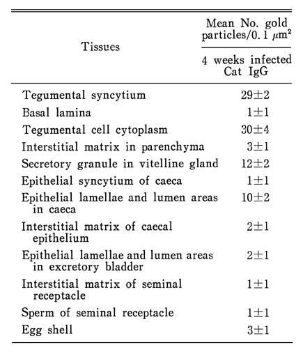
In order to determine the antigenic localization in the tissues of the adult Metagonimus yokogawai, immunogoldlabeling method was applied using serum immunoglobulins(IgG) of cats which were infected with isolated metacercariae from Plecoglossus altivelis. The sectioned worm tissue was embedded in Lowicryl HM 20 medium and stained with infected serum IgG and protein A gold complex(particle size: 12 nm). It was observed by electron microscopy at each tissue of the worm.
The gold particles were observed on the tegumental syncytium as well as cytoplasm of tegumental cells and epithelial lamella of the caecum. The gold particles were not observed on the basal lamina of the tegument, interstitial matrix of the parenchyma, the muscle tissue and mitochondria of the tegument.
The gold particles were specifically labeled in the secretory granules in the vitelline cells. They were also labeled on the lumen of bladder and egg shell.
The above findings showed that antigenic materials in the tissue of adult worms were specifically concentrated on the tegumental syncytium as well as cytoplasm of tegumental cells and epithelial lamella of the caecum.
Figures
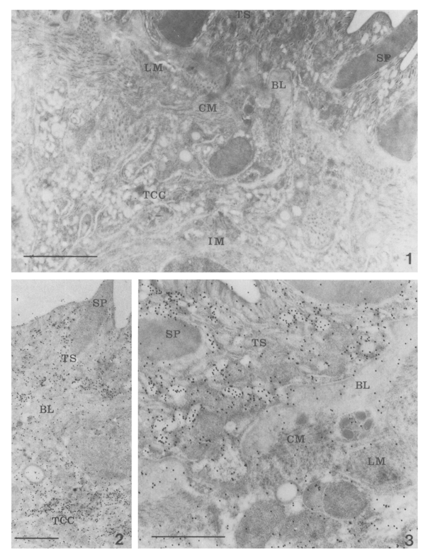
Figs. 1-3 Fig. 1. Electron micrographs of the tegument of the worm, which was reacted with cat IgG from noninfected control, showed the tegumental syncytium(TS), basal layer(BL), circular muscle(CM) layer, longitudinal muscle(LM) later, interstitial matrix(IM) and tegumental cell cytoplasm(TCC). Gold particles were not labeled on the tegument or other portions of the tissue. Bar=1 µm (×28,000)
Figs. 2, 3. The tegumental tissue of the worm reacted with specific antibody(IgG) from infected dog. Gold particles were specifically labeled in the tegumental syncytium and cytoplasm of the tegumental cell. Bar=1 µm (×17,000; ×28,000)
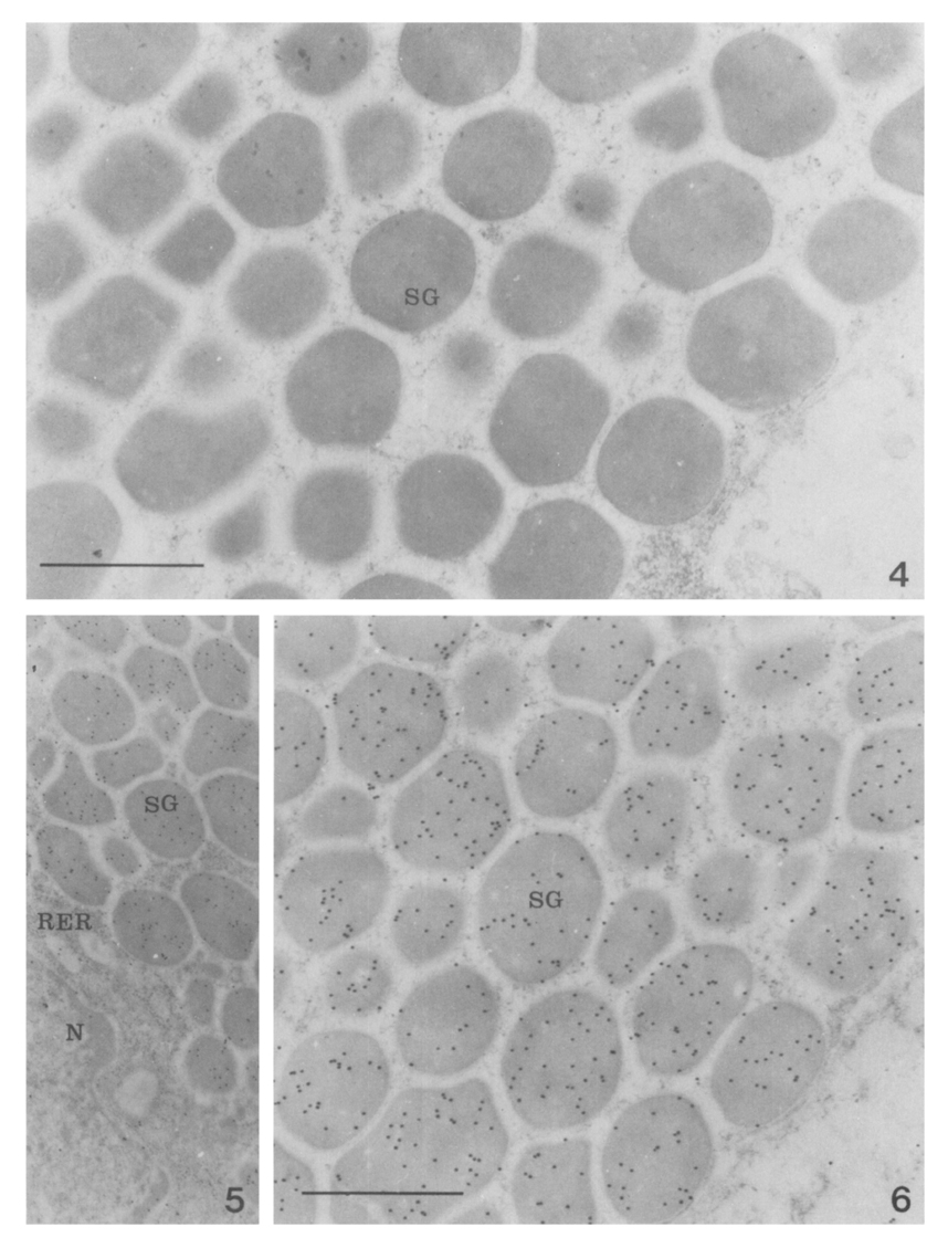
Figs. 4-6 Fig. 4. The vitelline gland of the worm which was reacted with cat IgG from noninfected control. Gold particles were not labeled on the cytop;asm and secretory granules of vitelline gland cell. Bar=1 µm (×28,000)
Figs. 5, 6. The vitelline gland of the worm which reacted with specific antibody(IgG) from infected cat. Gold particles were very specifically labeled on the secretory granules of vitelline gland cell cytoplasm. Bar=1 µm (×17,000; ×28,000)
Figs. 7-8 Fig. 7. The caecal section of the worm reacted with control group cat IgG from noninfected control. Gold particles were not labeled on the all area of the caecum. Bar=1 µm (×28,000)
Fig. 8. The caecal section of the worm reacted with specific antibody (IgG) from infected cat IgG. Gold particles were predominantly labeling on the caecum tegumental syncytium. Bar=1 µm (×28,000)
Figs. 9-11 Fig. 9. The sperms in the seminal receptacle of the worm which reacted with specific antibody(IgG) from infected cat. Gold particles were predominantly labeled on the tail part of the sperm and interstitial matrix of the seminal receptacle cell. Bar=1 µm (×28,000)
Fig. 10. The epithelium of excretory bladder of the worm reacted with control group dog IgG from noninfected control. Gold particles were predominantly labeled on the epithelial lamellae. Bar=1 µm (×17,000)
Fig. 11. The epithelium of excretory bladder of the worm which reacted with specific antibody(IgG) from infected cat. Gold particles were predominantly labeled on the epithelial lamellae. Bar=1 µm (×17,000)
Figs. 12-13 Fig. 12. The egg in the uterus of the worm reacted with control group cat IgG from noninfected control. Gold particles were not labeled on the egg shell. Bar=1 µm (×28,000)
Fig. 13. The egg in the uterus of the worm which reacted with specific antibody(IgG) from infected cat. Gold particles were predominantly labeled on the epithelial lamellae. Bar=1 µm (×28,000)
Tables
Table 1 Quantitative density of the labeled gold particles in the tissues of Metagonimus yokogawai reacted with antibody(IgG)* obtained from cats infected with Metagonimus yokogawai
References
1.
Chai JY. Seoul J Med 1979;20(2):104–117.
2.
Cho KM, et al. Yonsei Rep Trop Med 1976;7:26–37.
3.
de Water R, Fransen JA, Deelder AM. Ultrastructural localization of the circulating anodic antigen in the digestive tract of Schistosoma mansoni using monoclonal antibodies in an immunogold labeling procedure. Am J Trop Med Hyg 1986;35(3):549–558.
4.
Fujino T, et al. Jpn J Parasit 1989;38(5):263–270.
5.
Imai J. Trop Med 1979;21(2):45–55.
6.
Inatomi S, et al. Jap J Parasit 1968;17(6):456–460.
7.
Chu BD, Rim HJ, Kim SJ. [A study on the body fluid antigen of Clonorchis sinensis using immunogold labeling method]. Korean J Parasitol 1990;28(1):11–23.
8.
Joo KH, Ahn H, Chung MS, Rim HJ. Demonstration of species-specific and cross reactive components of Paragonimus westermani crude worm antigen by EITB. Korean J Parasitol 1989;27(1):9–14.
9.
Kang SY, Cho SY, Chai JY, Lee JB, Jang DH. A Study On Intestinal Lesions Of Experimentally Reinfected Dogs With Metagonimus Yokogawai. Korean J Parasitol 1983;21(1):58–73.
10.
Kim SI, Ko EK, Kang SY, Cho SY. [Antigenicity of the soluble egg antigen of Paragonimus westermani]. Korean J Parasitol 1986;24(1):49–54.
11.
Lee JB, Chi JG, Lee SK, Cho SY. Study On The Pathology Of Metagonimiasis In Experimentally Infected Cat Intestine. Korean J Parasitol 1981;19(2):109–129.
12.
Lee SH, Seo BS, Chai JY, Hong SJ. [Study on Metagonimus yokogawai(Katsurada, 1912) in Korea VII. Electron microscopic observation on the tegumental structure]. Korean J Parasitol 1984;22(1):1–10.
13.
Lee SH, Sung SH, Chai JY. [Immunohistochemical study on the antigenicity of body compartments of Paragonimus westermani]. Korean J Parasitol 1989;27(2):109–117.
14.
von Lichtenberg F, Bawden MP, Shealey SH. Origin of circulating antigen from the schistosome gut. An immunofluorescent study. Am J Trop Med Hyg 1974;23(6):1088–1091.
15.
Nash TE. Localization of the circulating antigen within the gut of Schistosoma mansoni. Am J Trop Med Hyg 1974;23(6):1085–1087.
16.
Ohnishi Y. Jpn J Parasit 1987;36(4):271–275.
17.
Ohnishi Y, et al. Jap J Vet Sci 1984;46:885–887.
18.
Roth J. The preparation of protein A-gold complexes with 3 nm and 15nm gold particles and their use in labelling multiple antigens on ultra-thin sections. Histochem J 1982;14(5):791–801.
19.
Roth J. Applications of immunocolloids in light microscopy. Preparation of protein A-silver and protein A-gold complexes and their application for localization of single and multiple antigens in paraffin sections. J Histochem Cytochem 1982;30(7):691–696.
20.
Saito S. Jap J Parasit 1972;21(6):449–458.
21.
Sugiyama H, Sugimoto M, Akasaka K, Horiuchi T, Tomimura T, Kozaki S. Characterization and localization of Paragonimus westermani antigen stimulating antibody formation in both the infected cat and rat. J Parasitol 1987;73(2):363–367.
22.
Sun T, et al. Jap J Med Sc Biol 1969;22:263–272.
23.
Yogore MG, et al. Am J Trop Med Hyg 1965;14(4):586–591.
24.
Yokogawa M, et al. Jap J Parasit 196;11(2):117–122.