Warning: mkdir(): Permission denied in /home/virtual/lib/view_data.php on line 81
Warning: fopen(upload/ip_log/ip_log_2024-04.txt): failed to open stream: No such file or directory in /home/virtual/lib/view_data.php on line 83
Warning: fwrite() expects parameter 1 to be resource, boolean given in /home/virtual/lib/view_data.php on line 84 Surface ultrastructure of Parvatrema timondavidi (Digenea:Gymnophallidae) according to its developmental stages
Surface ultrastructure of Parvatrema timondavidi (Digenea:Gymnophallidae) according to its developmental stages
Jae Ran Yu,*1Jin Young Park,2 and Jong Yil Chai2
1Department of parasitology, College of Medicine, Kon-Kuk University, Chungju 380-701, Korea.
2Department of Parasitology and Institute of Endemic Diseases, Seoul National University College of Medicine, Seoul 110-799, Korea.
Received April 28, 1994; Accepted May 28, 1994.
Abstract
Surface ultrastructure of Parvatrema developmental stages was studied using a scanning electron microscope. The metacercariae were collected from the marine clam, Tapes philippinarum, and juvenile and worms adult were recovered at 1, 2, 3, and 7 days after experimental infection of mice. The metacercariae had a large oral sucker and characteristic lateral projections. Around the lip of the oral sucker many type I and type II sensory papillae were observed, and type III papillae were located symmetrically on the medial side of the lateral projection. Numerous type I papillae were grouped around the genital pore. The tegumental spines were distributed over the worm surface except the lip of the sucker and genital pore. The 1-day old worm had a well-developed ventral sucker, with 6 type II sensory papillae on its outer surface and another 6 type I papillae on the inner side, Two small type I papillae were seen on the anterior side of the ventral sucker. The genital pore was and 15 type I papillae were grouped around it. The 2-, 3-, and 7-day worms revealed that as they grew to be adults, the spine tips became multipointed, the genital pore formed a genital atrium, and the cytoplasmic process became well differentiated. In 2- and 3-day worms 10 type II papillae encircling the lip of the oral sucker, and additional 4 papilled at the dorsal side of 4 dorsal type II papillae were a characteristic feature. The distribution pattern of sensory papillae around the oral sucker and genital pore, and 2 type I papillae on the anterior side of the ventra sucker, was so peculiar in P. timondavidi, that they seem to be useful keys for taxonomic differentiation from other gymnophallids.
Figures
Figs. 1-9 Scanning electron micrographs of the metacercaria of Parvatrema timondavidi. Fig. 1. Whole ventro-lateral view of a metacercaria. Bar = 16.7 µm. Fig. 2. Oral sucker showing type I papillae (open arrow), type II papillae (open arrow), and lateral projection (*). Bar = 5.0 µm. Fig. 3. Magnification of a lateral projection on the lip of the oral sucker of Fig. 2, showing a type III sensory papilla (*) on its medial side. Bar = 1.1 µm. Fig. 4. Magnification of the ventral sucker showing type II sensory papillae (arrow heads) on its lip portion. Bar = 1.7 µm. Fig. 5. Tegument nearby the genital pore showing many type I sensory papillae (arrow heads). Bar = 1.7 µm. Fig. 6. Tegument around the excretory pore (*). Bar = 1.7 µm. Fig. 7. Tegumental spines at the oral sucker level, with 4-5 pointed tips. Bar = 1.7 µm. Fig. 8. Spade-shaped tegumental spines at the ventral sucker level. Bar = 1.7 µm. Fig. 9. Tegumental spines at the excretory pore level, with 0-2 tips. Bar = 1.7 µm.
Figs. 10-16 Scanning electron micrographs of 1-day (Figs. 10-13) and 2-day old (Figs. 14-16) juveniles of P. timondavidi. Fig. 10. whole ventral view of a 1-day old worm. Bar = 14.3 µm. Fig. 11. Magnification of a lateral projection on the lip of the oral sucker, showing the smooth cytoplasmic membrance. Bar = 1.7 µm. Fig. 12. Genital pore (large arrows) and type I sensory papillae (small arrows). Bar = 1.0 µm. Fig. 13. Distribution of sensory papillae in the ventral sucker. An inner circle of type I papillae (white arrow heads), an outer circle of type II papillae (black arrows), and 2 type I papillae (*) ard characteristically distributed. Bar = 2.9 µm. Fig. 14. Whole ventral view of a 2-day old worm, showing oral, ventral suckers, and genital proe (arrows), Bar = 14.3 µm. Fig. 15. A lateral projection on the lip of the oral sucker, showing many cytoplasmic folds. Bar = 1.7 µm. Fig. 16. Tegumental spines at the ventral sucker level, with 6-7 pointed tips. Transverse cytoplasmic processes are also well developed. Bar = 1.0 µm.
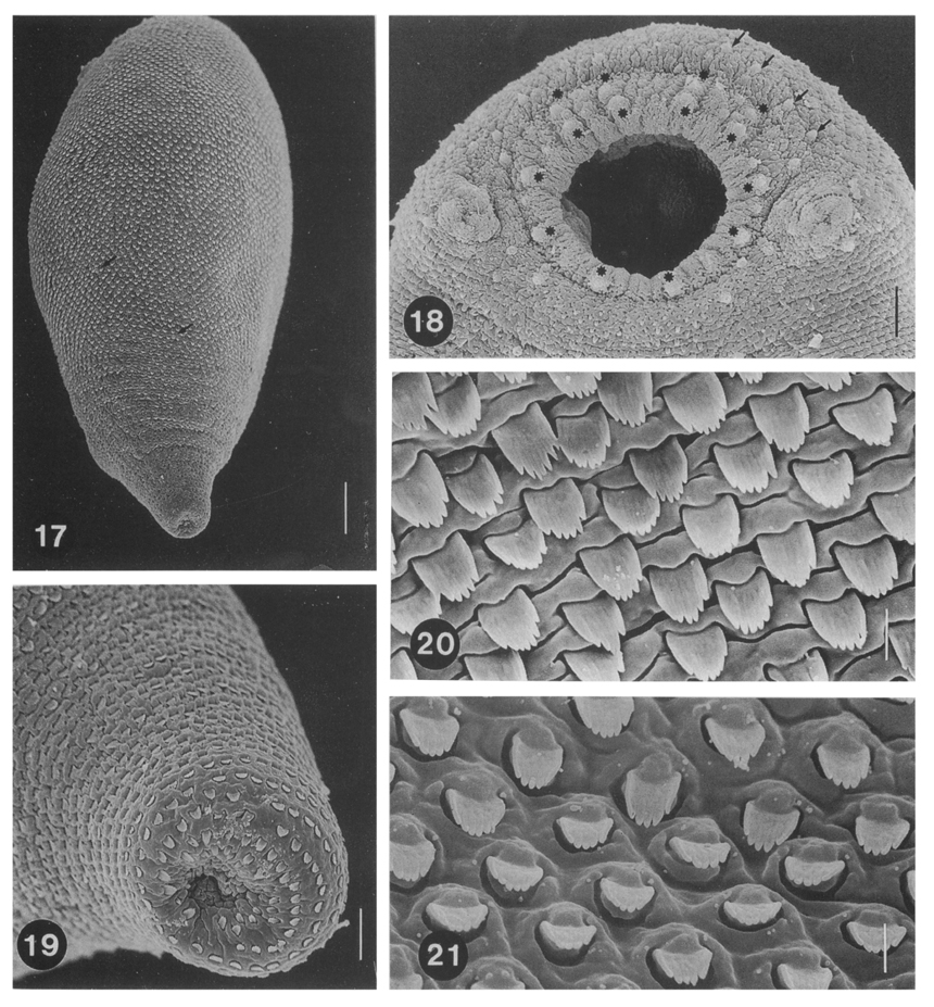
Figs. 17-21 Scanning electron micrographs of 3-day old juveniles of P. timondavidi. Fig. 17. Dorsal view of a 3-day old worm. Type I sensory papillae (arrows) are seen. Bar = 13.3 µm. Fig. 18. Oral sucker and two lateral projections. On the lip of the oral sucker 10 type II sensory papillae (*) are arranged almost equidistantly, of which anterior 4 papillae are overlapped anteriorly by 4 additional papillae, and half-circled by many type I papillae (arrows) bilaterally. Bar = 5.4 µm. Fig. 19. Excretory pore surrounded by tegumental spines. Bar = 3.9 µm. Fig. 20. Tegumental spines at the ventral sucker level. Transverse cytoplasmic folds are well developed, and the tip of the spines are divided into 6-7 points. Bar = 1.0 µm. Fig. 21. Tegumental spines at the excretory pore level with 5-6 pointed tips. The cytoplasmic membrane is folded as rhomboid shape. Bar = 1.0 µm.
Figs. 22-28 Scanning electron micrographs of 7-day old adult worms of P. timondavidi. Fig. 22. Whole ventro-lateral view of a 7-day old worm, showing two suckers and a genital atrium (*). Bar = 16.7 µm. Fig. 23. Type I (white *) and type II (back *) sensory papillae around the oral sucker. Bar = 1.2 µm. Fig. 24. A spineless area around the genital pore. Bar = 3.3 µm. Fig. 25. Magnification of the genital pore showing many type I sensory papillae. Bar = 1.0 µm. Fig. 26. Tegumental spines at the oral sucker level. Transverse cytoplasmic folds are well developed, and the spines are multipointed. Bar = 1.0 µm. Fig. 27. Tegumental spines at the ventral sucker level. Longitudinal folds are produced. Bar = 1.0 µm. Fig. 28. Tegumental spines at the excretory pore level. Cytoplasmic folds are rectangular shape. Bar = 1.0 µm.
References
1.
Lee SH, Seo BS, Chai JY, Hong SJ. [Study on Metagonimus yokogawai(Katsurada, 1912) in Korea VII. Electron microscopic observation on the tegumental structure]. Korean J Parasitol 1984;22(1):1–10.
2.
Lee SH, Hong SJ, Chai JY, Hong ST, Seo BS. [Tegumental ultrastructures of Echinostoma hortense observed by scanning electron microscopy]. Korean J Parasitol 1986;24(1):63–70.
3.
Lee SH, Kim SJ, Chai JY, Sohn WM. [Tegumental ultrastructures of Paragonimus iloktsuenensis according to the developmental stages]. Korean J Parasitol 1989;27(1):57–66.
4.
Seo BS, Lee SH, Chai JY, Hong ST, Hong SJ. [Studies on intestinal trematodes in Korea X. Scanning electron microscopic observation on the tegument of Fibricola seoulensis]. Korean J Parasitol 1984;22(1):21–29.
6.
Bartoli P. Comptes rendus des seances de l'Academie des sciences. Serie, Paris 1963;257(2):518–520.
7.
Bennett CE. Scanning electron microscopy of Fasciola hepatica L. during growth and maturation in the mouse. J Parasitol 1975;61(5):892–898.
Hong SJ, Chai JY, Lee SH. Surface ultrastructure of the developmental stages of Heterophyopsis continua (Trematoda: Heterophyidae). J Parasitol 1991;77(4):613–620.
10.
Lee SH, Chai JY, Hong ST. Gymnophalloides seoi n. sp. (Digenea: Gymnophallidae), the first report of human infection by a gymnophallid. J Parasitol 1993;79(5):677–680.
12.
Lumsden RD. Surface ultrastructure and cytochemistry of parasitic helminths. Exp Parasitol 1975;37(2):267–339.
13.
Pekkarinen M. Ann Zool Fennici 1984;21:481–498.
14.
Pekkarinen M. Ann Zool Fennici 1987;24:29–37.
15.
Yu JR, Chai JY, Lee SH. Parvatrema timondavidi (Digenea; Gymnophallidae) transmitted by a clam, Tapes philippinarum, in Korea. Korean J Parasitol 1993;31(1):7–12.