Warning: mkdir(): Permission denied in /home/virtual/lib/view_data.php on line 81

Warning: fopen(upload/ip_log/ip_log_2024-04.txt): failed to open stream: No such file or directory in /home/virtual/lib/view_data.php on line 83

Warning: fwrite() expects parameter 1 to be resource, boolean given in /home/virtual/lib/view_data.php on line 84
Eimeria pipistrellus n. sp. from Pipistrellus kuhlii (Chiroptera: Vespertilionidae) in Saudi Arabia
| Home | E-Submission | Sitemap | Contact us |  
top_img
Korean J Parasito Search

CLOSE

Korean J Parasito > Volume 37(1):1999 > Article
Alyousif, Al-Dakhil, and Al-Shawa: Eimeria pipistrellus n. sp. from Pipistrellus kuhlii (Chiroptera: Vespertilionidae) in Saudi Arabia

Abstract

Fecal samples from 12 Pipistrellus kuhlii captured at Shagrah, Saudi Arabia, were examined for coccidia and three (25%) found to harbor a undescribed eimerian, herein described as Eimeria pipistrellus n. sp. Sporulated oocysts were subspherical, 24.8×23.2 (22-27×20-25) µm, with a bilayered and smooth wall. The micropyle was absent, but a large oocyst residuum and a single polar granule were present. Sporocysts were ovoid, 11.6×8.3 (10.5-13×7.5-9) µm, with a prominent Stieda body, but without a substiedal body; sporozoites lay head to tail in sporocysts and contained one large posterior refractile body. Eimeria pipistrellus n. sp. is the 3rd species of the genus Eimeria found from bats of the genus Pipistrellus.

INTRODUCTION

To date 17 eimerian species have been described from chiropterans worldwide (Duszynski and Barkley, 1985; Duszynski et al., 1988; Duszynski, 1997; Scott and Duszynski, 1997), but only two species of Eimeria have been reported from members of the genus Pipistrellus; E. macyi from Pipistrellus subflavus from USA (Wheat, 1975) and E. redukeri from P. javanicus from Japan (Duszynski, 1997).
No Eimeria species has been described from Pipistrellus kuhlii, herein we describe a new species of Eimeria from P. kuhlii from Shagrah, Saudi Arabia.

MATERIALS AND METHODS

Twelve bats, P. kuhlii (Natterer, 1819) were trapped alive at Shagrah during November, 1997 with the aid of mist nets. All bats were killed in the laboratory, and the abdominal cavity was opened. The intestinal tract, caecum, rectum and colon were slit lengthwise, and their contents were collected separately and mixed with 2.5% (w/v) aqueous potassium dichromate solution to inhibit bacterial growth. The suspension was then spread in a thin layer in petri dishes and incubated at 26 ± 2℃ for 1 week so that the oocysts could sporulate. The preparations were then microscopically examined for sporulation at 12 hr intervals. Sporulated oocysts were concentrated by flotation with Sheather' sugar solution (McAllister et al., 1995). Fifty sporulated oocysts and 50 sporocysts were examined, and measured with a microscope fitted with a ×100 apochromatic oil immersion objective and a ×10 ocular micrometer. The number of layers of the oocyst wall, its thickness, and detailed structure of the sporocysts were examined after crushing the oocysts by pressure on the coverslip. All measurements are in micrometers (µm) with the mean followed by the range in parentheses.

RESULTS

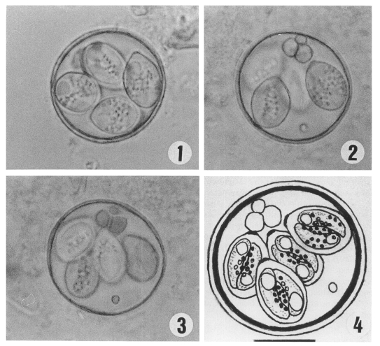
Eimeria pipistrellus (Figs. 1-4) Description

Oocyst wall 1.2-1.5 (1.3) thick consisting of 2 layers of approximately equal thickness; outer, light brownish-yellow, smooth; inner dark, smooth; a micropyle is absent, but an oocyst residuum, consisting of one to three large globules, 5.2 (4.5-6.0), and a polar granule approximately 1.6 both are present. Sporulated oocysts (n=50) subspheroidal 24.8×23.2 (22-27×20-25) with L:W ratio 1.0-1.2 (1.06). Sporocysts ovoidal, slightly pointed at one end, 11.6×8.3 (10.5-13×7.5-9) with L:W ratio 1.4-1.5 (1.45), Stieda body present, but sub- and parastieda bodies absent. Sporocyst residuum present as numerous minute dispersed granules. Sporozoites elongate, lying head to tail, each with one large posterior refractile body.

Taxonomic summary

Type host: Pipistrellus kuhlii (Kuhl, 1819) Vespertilionidae
Type locality: Shagrah, central region, Saudi Arabia
Prevalence: Found in three of twelve (25%) P. kuhlii.
Site of infection: Unknown, oocysts recovered from intestinal contents.
Sporulation time: Six days at 26 ± 2℃
Etymology: The specific name Pipistrellus is derived from the generic name of the host.
Type specimens: Oocysts in 10% formalin and a phototype are deposited in the Parasitological Collection, Department of Zoology, College of Science, King Saud University, Riyadh, Saudi Arabia both as KSUC 105.

DISCUSSION

Only two species of Eimeria have been described previously from the genus Pipistrellus (Vespertilionidae), E. macyi (Wheat, 1975) and E. redukeri (Duszynski, 1997). Eimeria pipistrellus n. sp. differs considerably from both of the above mentioned two species in having a larger oocyst with smooth outer wall. Moreover, E. pipistrellus can be distinguished easily from E. macyi because it has an oocyst residuum and lacks a substiedal body, and from E. redukeri in having a larger oocyst residuum composed of one to three large globules, in having larger sporocysts with smaller length/width ratio, and in having small, dispersed granules as a sporocyst residuum rather than only one to three large globules seen in the sporocysts of E. redukeri.
These differences in structural features, geographic distribution, and host species make us suggest that E. pipistrellus is a distinct form.

ACKNOWLEDGEMENTS

We wish to thank Dr. Awad El-Bahrawy for his great help in collecting the bats.

REFERENCES

1. Duszynski DW. Coccidia from bats (Chiroptera) of the world: A new Eimeria species in Pipistrellus javanicus from Japan. J Parasitol 1997;83:280-282. PMID: 9105311.
crossref pmid
2. Duszynski DW, Barkley LJ. Eimeria from bats of the world: A new species in Tomopeas ravus from Peru. J Parasitol 1985;71:204-208. PMID: 3998959.
crossref pmid
3. Duszynski DW, Reduker DW, Parker BB. Eimeria from bats of the world. II. A new species in Tadarida femorasacca from Sonora, Mexico. J Parasitol 1988;74:317-321. PMID: 3357122.
crossref pmid
4. McAllister CT, Upton SJ, Jacobson ER, Kopit W. A description of Isospora amphiboluri (Apicomplexa: Eimeriidae) from the inland bearded dragon, Pogona vitticeps (Sauria: Agamidae). J Parasitol 1995;81:281-284. PMID: 7707208.
crossref pmid
5. Scott DT, Duszynski DW. Eimeria from bats of the world; two new species from Myotis spp. (Chiroptera: Vespertilionidae). J Parasitol 1997;83:495-501. PMID: 9194833.
crossref pmid
6. Wheat BE. Eimeria macyi sp.n. (Protozoa: Eimeriidae) from the eastern pipistrelle, Pipistrellus subflavus from Alabama. J Parasitol 1975;61:920-922. PMID: 1185436.
crossref pmid
Figs. 1-4
Photomicrographs and an drawing of sporulated oocysts of Eimeria pipistrellus from naturally infected Pipistrellus kuhlii (×1,000).
Fig. 1. The Stieda body and refractile globules. Fig. 2. The oocyst and sporocyst residuum. Fig. 3. The spherical, small polar granule. Fig. 4. A camera lucida drawing of sporulated oocyst of E. pipistrellus. Scale bar = 10 µm.
kjp-37-1-g001.jpg
Table 1.
Comparative data of Eimeria species from pipistrelle bats
E. macyi E. redukeri E. pipistrellus
Oocyst
 shape subspherical to subspherical subspherical
 size (μm) broadly ellipsoid
16-21 × 15-19 16-25 × 14-21 22.3-27.2 × 20-25.4
(19 × 17.6) (20.3 × 18.1) (24.8 × 23.2)
 wall rough mammiliated smooth
 micopyle absent absent absent
 residuum bsent single (2.0 × 3.8) one to three
5.2 (4.5-5.6)
 polar granule ellipsoid ellipsoid spheroid
Sporocyst
 shape ovoid ovoid ovoid
 size (μm) 10-12 × 6-8 8-12 × 5-8 10.5-12.9×7.5-9.4
(11 × 7) (10.6 × 6.6) (11.6 × 8.3)
 residuum several granules 1-3 globules several granules
Host Pipistrellus subflavus Pipistrellus javanicus Pipistrellus kuhlii
Locality USA Japan Saudi Arabia
Citation Wheat (1975) Duszynski (1997) Present study
TOOLS
PDF Links  PDF Links
PubReader  PubReader
ePub Link  ePub Link
XML Download  XML Download
Full text via DOI  Full text via DOI
Download Citation  Download Citation
  Print
Share:      
METRICS
3
Crossref
5
Scopus
5,821
View
75
Download
Editorial Office
Department of Molecular Parasitology, Samsung Medical Center, School of Medicine, Sungkyunkwan University,
2066 Seobu-ro, Jangan-gu, Suwon 16419, Gyeonggi-do, Korea.
Tel: +82-31-299-6251   FAX: +82-1-299-6269   E-mail: kjp.editor@gmail.com
About |  Browse Articles |  Current Issue |  For Authors and Reviewers
Copyright © 2024 by The Korean Society for Parasitology and Tropical Medicine.     Developed in M2PI