Anisakiasis is one of the fish-transmitted infection that results from accidental ingestion of third-stage larvae belonging to the family Anisakidae. Human infection can occur by consumption of raw or undercooked fish, such as mackerel, squid, cod, anchovy, salmon and tuna. These fishes act as an intermediate host for ascarids of sea mammals. About two thirds of people who eat these fishes harboring larva experienced gastric or intestinal symptoms, including epigastric pain, nausea, vomiting and etc., two to six hours afterwards (Sugimachi et al., 1985). Many reports have been issued on gastric and intestinal anisakiasis in Korea (Im et al., 1990, 1995) and elsewhere (Muraoka et al., 1996; Lopez-Serrano et al. 2000), but most of the cases have involved a single worm infection (Kim et al., 1991). Although, Seol et al. (1994) reported upon the removal of twenty-two worms from 20 Korean patients, they did not specify the number of worms collected from each patient. We have experienced a case of multiple infection of anisakid larvae in one patient.
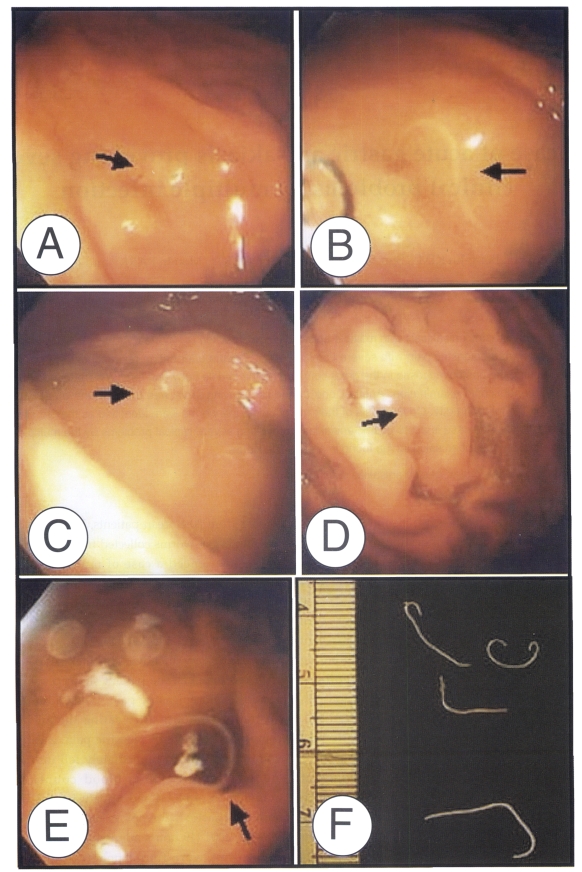
A 68-year-old Korean woman, residing in Busan, Korea, was admitted to the emergency room at Kosin University Hospital in May 12, 2002. About one hour after eating raw anchovies in thick soypaste mixed with red peppers, the patient showed symptoms of vomiting and epigastric pain. When admitted to the hospital five hours after eating the anchovy, she complained of severe epigastric pain, chest pain and vomiting, and then immediately fell into a state of mental stupor. A total blood count, chemical examination of blood and urinalysis, and EKG were normal. A gastroendoscopic examination was done on May 13, 2002 (19 hours admission), and four thread-like worms were found penetrating the gastric mucosa in the great curvature of the middle body and fundus (Figs. 1A to 1D). Another one long-white nematode was found at the great curvature side of upper body during an endoscopic examination performed on the following day (Fig. 1E). The gastric mucosa of the body and antrum showed diffuse hyperemia. No more worms could be found during a third endoscopic examination carried out on two days after the initial examination. The state of patient improved immediately after removing the worms and she was discharged after receiving supportive care on 17 May 2002.
The worms were fixed in 10% formalin, cleared in glycerin-alcohol, mounted with glycerin-jelly and observed under a light microscope equipped with a micrometer. The length and maximum- width of the worms were as follows; 16.5 × 0.3 mm, 28.1 × 0.5 mm, 19.5 × 0.4 mm, and 17.5 × 0.3 mm (Fig. 1F). The fifth worm extracted from the second endoscopic examination was not secured. The third-stage larvae usually have average length of 20.0 - 30.0 mm, and slender esophagus is followed by ventriculus which forms an oblique junction at the posterior end with the intestine. The larvae have lips at the anterior end and a mucron at the posterior end. The worms of this report had mouths equipped with three lip bulges and boring teeth, a mucron at the posterior part. We were unable to find an intestinal cecum. The measurements and indices of the present specimens (Table 1) also represent common characteristics and morphological features of the third-stage larvae of Anisakis simplex. On the basis of the morphology and measurements, the parasites were identified as the third-stage larvae of Anisakis simplex.
Four species of anisakid are known, i.e. A. simplex type I and type II, and a Pseudoterranova type A and Contracaecum sp. Most anisakiasis, however, take place by the larva of Anisakis simplex. The main sources of anisakid infection in Korea are, cuttle-fish, yellow corvina, sea eel, ling, and yellowtail (Im et al., 1995). Song et al. (1995) reported that the muscle of anchovy is highly infected with anisakid larvae. Moreover, their report (Song et al., 1995) suggests that human infection is possible from the consumption of anchovy. This case confirms that anchovies can cause anisakiasis. Most reports on anisakiasis indicate that the most frequent parasitic location is in the great curvature of the lower, mid and upper body. In the present case worms were also distributed over the great curvature of the fundus, the middle body and the upper body. The reason why the worms are recovered primarily from the great curvature is not clear. Shibata et al. (1989) noted that the great curvature provides a good environment for anisakid larvae for penetrating the gastric wall, due to the large number of folds and the more active secretion of mucus in this region.
Although Choi et al. (1989) reported finding two larvae in one patient among five anisakiasis cases in Korea, almost all anisakiasis cases have recorded single worm removal in one patient. We found four larvae during the first gastroendoscopic examination and another one in a second examination. The patient in this case experienced typical gastric anisakiasis symptoms and fell into mentor stupor by the intolerable epigastric pain. Therefore, we recommend that multiple anisakid larval infection should be borne in mind.