Warning: mkdir(): Permission denied in /home/virtual/lib/view_data.php on line 81

Warning: fopen(upload/ip_log/ip_log_2024-04.txt): failed to open stream: No such file or directory in /home/virtual/lib/view_data.php on line 83

Warning: fwrite() expects parameter 1 to be resource, boolean given in /home/virtual/lib/view_data.php on line 84
Short-Cut Pathway to Synthesize Cellulose of Encysting Acanthamoeba
| Home | E-Submission | Sitemap | Contact us |  
top_img
Korean J Parasito Search

CLOSE

Korean J Parasito > Volume 50(4):2012 > Article
Moon and Kong: Short-Cut Pathway to Synthesize Cellulose of Encysting Acanthamoeba

Abstract

The mature cyst of Acanthamoeba is highly resistant to various antibiotics and therapeutic agents. Cyst wall of Acanthamoeba are composed of cellulose, acid-resistant proteins, lipids, and unidentified materials. Because cellulose is one of the primary components of the inner cyst wall, cellulose synthesis is essential to the process of cyst formation in Acanthamoeba. In this study, we hypothesized the key and short-step process in synthesis of cellulose from glycogen in encysting Acanthamoeba castellanii, and confirmed it by comparing the expression pattern of enzymes involving glycogenolysis and cellulose synthesis. The genes of 3 enzymes, glycogen phosphorylase, UDP-glucose pyrophosphorylase, and cellulose synthase, which are involved in the cellulose synthesis, were expressed high at the 1st and 2nd day of encystation. However, the phosphoglucomutase that facilitates the interconversion of glucose 1-phosphate and glucose 6-phosphate expressed low during encystation. This report identified the short-cut pathway of cellulose synthesis required for construction of the cyst wall during the encystation process in Acanthamoeba. This study provides important information to understand cyst wall formation in encysting Acanthamoeba.

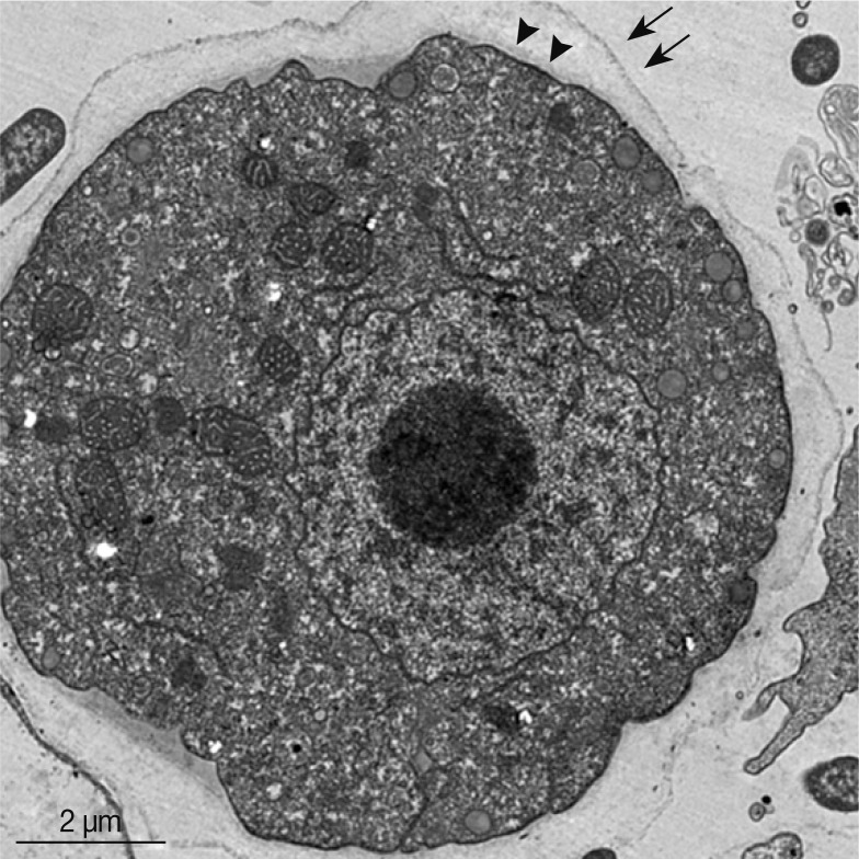
Under unfavorable environmental conditions, Acanthamoeba trophozoites transform into cysts those are resistant to extreme physical and chemical conditions [1]. The mature cyst has 2 walls, an outer wall (exo-cyst) and an inner wall (endo-cyst) (Fig. 1). The cyst wall of Acanthamoeba castellanii contains carbohydrates (35%), protein (33%), ash (8%), lipid (4-6%), and unidentified materials (20%) [2]. Acid-resistant proteins and cellulose are the major components of the cyst wall [3]. The precursor of cellulose is glucose, and the source of glucose is glycogen in encysting amoeba [4]. According to the previous report, the glycogen molecule undergoes rapid degradation during the early phase of Acanthamoeba encystation, and the glycogen content of cysts is significantly less (18 µg/106 cells) than that of trophozoites (83 µg/106 cells) [4]. These results suggest the involvement of glycogen metabolism in cellulose synthesis during encystation of Acanthamoeba. However, the metabolic pathway of glycogen breakdown and cellulose synthesis during encystation has yet to be clarified. In this study, we hypothesized that the short-cut process of cellulose synthesis could be involved in rapid construction of the cyst wall of encysting Acanthamoeba. To conduct this study, A. castellanii Castellani (ATCC no. 30011) was used for induction of cysts, and comparison of ESTs and microarray analysis [7,8]. Real-time PCR analysis was performed to compare the expression levels of 3 enzymes involved in glycogen degradation and cellulose synthesis. Three sets of primers were used (Table 1), and 18s rDNA was used as a reference gene [8].
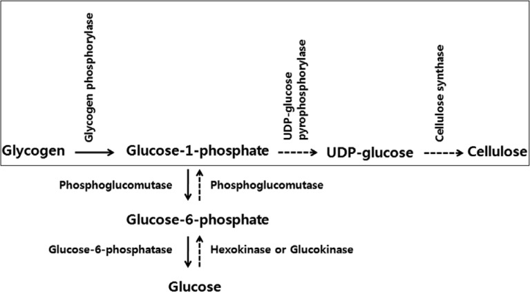
Fig. 2 shows the metabolic pathway of glycogen degradation (arrow pathway) and cellulose synthesis (dot-arrow pathway). In general, breakdown of glycogen into units of glucose occurs through phosphorylitic cleavage by glycogen phosphorylase, phosphoglucomutase, and glucose-6-phosphatase [5]. Synthesis of cellulose from glucose is also a multi-step process involving 4 enzymes; hexokinase, phosphoglucomutase, UDP-glucose pyrophosphorylase, and cellulose synthase [6]. According to the process, it is 7 steps from glycogen to cellulose in Acanthamoeba. Glucose-1-phosphate is a metabolic product of glycogen in glycogenolysis and converts to glucose through glucose-6-phosphate. For cellulose synthesis, glucose-1-phosphate is also an important branch point to generate cellulose from glucose (Fig. 2). Therefore, we hypothesized that the short-cut process of cellular synthesis from glycogen could be mediated by 3 enzymes in Acanthamoeba (Fig. 2, box).
Our previous ESTs analysis study provided partial sequences of glycogen phosphorylase (GenBank no. JX312797) and UDP-glucose pyrophosphorylase (GenBank no. JX312798) [7]. Using sequence information on cellulose synthase from A. castellanii Neff (Tang et al., unpublished), we cloned a partial cDNA of cellulose synthase from A. castellanii Castellani (GenBank no. JX312799). Results of cDNA microarray analysis between cysts and trophozoites indicated a 2.44-fold higher expression of glycogen phosphorylase in cysts than in trophozoites [8]. However, phosphoglucomutase was expressed less in cysts than in trophozoites (2.4-fold) [8]. These results suggested that the conversion of glucose-1-phosphate to glucose-6-phosphate is not essential in glycogen degradation during encystation of Acanthamoeba. Then, it is possible to hypothesize the short process to synthesize cellulose from glycogen through glucose-1-phosphate, an important branch point.
The results of the quantitative real-time PCR analysis of expression of 3 enzymes supported our hypothesis strongly. As shown in Fig. 3, the mRNA level of glycogen phosphorylase showed a gradual increase and reached the maximum (10.3-fold) at the third day after induction of encystation (Fig. 3A). Dictyostelium discoideum, a species of soil-living amoeba, has been reported to have 2 forms of glycogen phosphorylase [9]. The 2 forms of the enzyme may play different roles in the development of Dictyostelium because they have different expression patterns. Glycogen phosphorylase 1 was found to be functional during differentiation of Dictyostelium into spores which have walls containing cellulose [10]. During the time course of development of Dictyostelium, the 'b' form (glycogen phosphorylase 2) showed a decrease, whereas the 'a' form (glycogen phosphorylase 1) showed an increase [11]. The partial glycogen phosphorylase of A. castellanii Castellani showed 65% similarity with glycogen phosphorylase 1 of D. discoideum, and showed high expression during encystation. The mRNA levels of UDP-glucose pyrophosphorylase of Acanthamoeba showed the highest expression (6.1-fold) on the second day after induction of encystation (Fig. 3B). UDP-glucose pyrophosphorylase 2 of D. discoideum was required for the differentiation and development of the amoeba [12]. The partial UDP-glucose pyrophosphorylase of A. castellanii showed 62% similarity with UDP-glucose pyrophosphorylase 2 of D. discoideum. The expression level of cellulose synthase of A. castellanii showed an increase (3.0-fold) on the second day after induction of encystation (Fig. 3C). The partial sequence of cellulose synthase of A. castellanii showed 99% similarity with that of A. castellanii Neff (ACC008015- Tang et al., unpublished) and 64% similarity with that of D. discoideum. These results suggest the involvement of these enzymes in the encystation process of Acanthamoeba.
The development of the acid-insoluble protein containing ectocyst wall and the cellulose containing endocyst wall leads to emergence of resistance to biocides in encysted Acanthamoeba [13]. Cellulose, the primary component of the cyst wall, is also used for diagnosis of Acanthamoeba cysts [14]. This is the first study that we identified 3 enzymes involving cellulose synthesis in Acanthamoeba and confirm the short-cut pathway to synthesize cellulose by analysis of their expression patterns during encystation. Information on cellulose synthesis is important to understand the mechanism of encystation and to diagnose cyst-forming protozoa. This key process of cellulose synthesis may aid in understanding of cyst wall formation in Acanthamoeba.

ACKNOWLEDGMENTS

This research was supported by the Kyungpook National University Research Fund, 2010 and the Brain Korea 21 Project in 2011. The authors wish to thank Professor Petrus Tang (Molecular Medicine Research Center, Chang Gung University, Taiwan) for providing the sequence information on cellulose synthase from Acanthamoeba castellanii Neff.

REFERENCES

1. Marciano-Cabral F, Cabral G. Acanthamoeba spp. as agents of disease in humans. Clin Microbiol Rev 2003;16:273-307. PMID: 12692099.
crossref pmid pmc
2. Neff RJ, Neff RH. The biochemistry of amoebic encystment. Symp Soc Exp Biol 1969;23:51-81. PMID: 4243920.
pmid
3. Tomlinson G, Jones EA. Isolation of cellulose from the cyst wall of a soil amoeba. Biochim Biophys Acta 1962;63:194-200. PMID: 13985444.
crossref pmid
4. Bowers B, Korn ED. The fine structure of Acanthamoeba castellanii (Neff strain). II. Encystment. J Cell Biol 1969;41:786-805. PMID: 5768875.
crossref pmid pmc
5. Greenberg CC, Jurczak MJ, Danos AM, Brady MJ. Glycogen branches out: New perspectives on the role of glycogen metabolism in the integration of metabolic pathways. Am J Physiol Endocrinol Metab 2006;291:E1-E8. PMID: 16478770.
crossref
6. Delmer DP, Amor Y. Cellulose biosynthesis. Plant Cell 1995;7:987-1000. PMID: 7640530.
crossref pmid pmc
7. Moon EK, Chung DI, Hong Y, Kong HH. Expression levels of encystation mediating factors in fresh strain of Acanthamoeba castellanii cyst ESTs. Exp Parasitol 2011;127:811-816. PMID: 21276446.
crossref pmid
8. Moon EK, Xuan YH, Chung DI, Hong Y, Kong HH. Microarray analysis of differentially expressed genes between cysts and trophozoites of Acanthamoeba castellanii. Korean J Parasitol 2011;49:341-347. PMID: 22355200.
crossref pmid pmc
9. Rutherford CL, Cloutier MJ. Identification of two forms of glycogen phosphorylase in Dictyostelium. Arch Biochem Biophys 1986;250:435-439. PMID: 3490829.
crossref pmid
10. West CM, Erdos GW. Formation of the Dictyostelium spore coat. Dev Genet 1990;11:492-506. PMID: 2096020.
crossref pmid
11. Cloutier MJ, Rutherford CL. Glycogen phosphorylase in Dictyostelium. Developmental regulation of two forms and their physical and kinetic properties. J Biol Chem 1987;262:9486-9493. PMID: 3110159.

12. Bishop JD, Moon BC, Harrow F, Ratner D, Gomer RH, Dottin RP, Brazill DT. A second UDP-glucose pyrophosphorylase is required for differentiation and development in Dictyostelium discoideum. J Biol Chem 2002;277:32430-32437. PMID: 12060658.
crossref
13. Turner NA, Russell AD, Furr JR, Lloyd D. Emergence of resistance to biocides during differentiation of Acanthamoeba castellanii. J Antimicrob Chemother 2000;46:27-34. PMID: 10882685.
crossref pmid
14. Derda M, Hadaś E. Use of the presence of cellulose in cellular wall of Acanthamoeba cysts for diagnostic purposes. Wiad Parazytol 2009;55:47-51. PMID: 19579785.
pmid
Fig. 1
Cross section of an encysting Acanthamoeba after 24 hr of encystment. The cyst wall consists of 2 layers, exo-cyst (arrows) and endo-cyst (arrowheads).
kjp-50-361-g001.jpg
Fig. 2
Glycolysis and cellulose biosynthesis pathway in Acanthamoeba. Acanthamoeba trophozoites synthesize glycogen from glucose, which later breakdown during encystation to generate cellulose. It takes 7 steps from glycogen to cellulose through glucose. We hypothesized a brief pathway of cellulose synthesis from glycogen in Acanthamoeba (box).
kjp-50-361-g002.jpg
Fig. 3
Real-time PCR analysis of 3 enzymes during encystation. (A) An increase in levels of expression of glycogen phosphorylase was observed on Day 1 (2.4-fold), Day 2 (6.7-fold), and Day 3 (10.3-fold). (B) High expression of UDP-glucose pyrophosphorylase on Day 1 (2.3-fold), Day 2 (6.1-fold), and Day 3 (4.0-fold). (C) Cellulose synthesis showed high levels of expression on Day 1 (2.8-fold), Day 2 (3.0-fold), and Day 3 (2.2-fold).
kjp-50-361-g003.jpg
Table 1.
Primer sequences used in real-time PCR analysis
Gene name Primer sequence
Glycogen phosphorylase F´-TCGAGGTTGCTGATGGTGTTC
R´-ACGCACAAGCACTTTGAGGAG
UDP-glucose pyrophosphorylase F´-ACAAACCACACAATGGCCACC
R´-TGGTCTTGTGAGCCGCTAACT
Cellulose synthase F´-TCATCTACATGTTCTGCGCCC
R´-CGATCCAGTTGTTGAGCATGC
Editorial Office
Department of Molecular Parasitology, Samsung Medical Center, School of Medicine, Sungkyunkwan University,
2066 Seobu-ro, Jangan-gu, Suwon 16419, Gyeonggi-do, Korea.
Tel: +82-31-299-6251   FAX: +82-1-299-6269   E-mail: kjp.editor@gmail.com
About |  Browse Articles |  Current Issue |  For Authors and Reviewers
Copyright © 2024 by The Korean Society for Parasitology and Tropical Medicine.     Developed in M2PI