Warning: mkdir(): Permission denied in /home/virtual/lib/view_data.php on line 81
Warning: fopen(upload/ip_log/ip_log_2024-04.txt): failed to open stream: No such file or directory in /home/virtual/lib/view_data.php on line 83
Warning: fwrite() expects parameter 1 to be resource, boolean given in /home/virtual/lib/view_data.php on line 84 A human case infected by the larva of Terranova type A in Korea
A human case infected by the larva of Terranova type A in Korea
Byong-Seol Seo,Jong-Yil Chai,Soon-Hyung Lee,Sung-Tae Hong,Jeong-Wook Seo and Sung-Hoon Noh
Department of Parasitology and Institute of Endemic Diseases, College of Medicine,Seoul National University, Seoul 110, Korea.
Department of General Surgery, Seoul District Armed Forces General Hospital, Korea.
Abstract
A human case infected with Terranova type A larva was found in Korea. The patient was a 23-year old soldier of the Korean Army and the chief complaint was acute abdominal pain. The pain was chiefly at right lower quadrant. Appendectomy was performed under the clinical impression of acute appendicitis. However, during the surgery, a nematode larva was found moving on the serosal surface of terminal ileum.
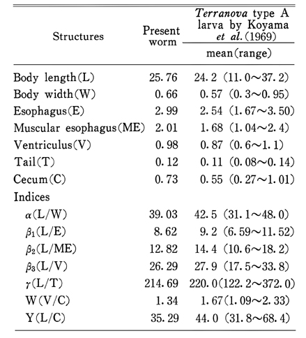
The worm was 25.76mm long and 0.66mm wide, and had the intestinal cecum reaching to anterior one-third level of ventriculus and a mucron at posterior end. Therefore, it was diagnosed as Terranova type A larva. This is the first human case of Terranova type A larva infection in Korea.
Figures
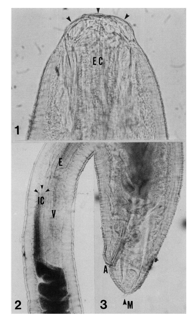
EXPLANATIONs FOR FIGURES Fig. 1. Head part of Terranova type A larva showing 3 lips (arrows) and excretory canal (EC), ×200.
Fig. 2. Muscular esophagus (E), ventriculus (V), intestine and intestinal cecum (IC) reaching over mid-level of ventriculus and demarcated by arrows. The characteristic arrangement of Terranova type A larva, ×40.
Fig. 3. Tail of Terranova type a larva with anus (A) and mucron (M), ×100.
Tables
Table 1 Measurements (mm) and indices of the anisakid larva from the present case in comparison with Terranova type A larva
References
1.
Cho SY, et al. Seoul J Med 1971;21(2):208–208.
2.
Fujino T, et al. Japanese J Parasitol 1984;33(2):73–92.
3.
Jeong JS, et al. Inje Med J 1984;5(3):359–367.
4.
Kagei N, et al. Japanese J Parasitol 1972;21(4):262–265.
5.
Kim CH, Chung BS, Moon YI, Chun SH. [A case report on human infection with Anisakis sp. in Korea]. Korean J Parasitol 1971;9(1):39–43.
6.
Koyama T, et al. Japanese J Parasitol 1969;18(5):466–487.
7.
Koyama T, et al. Japanese J Parasitol 1972;21(4):257–261.
8.
Lee KH, et al. Korean J Int Med 1981;24(12):1220–1227.
9.
Nagano K, et al. Nippon Iji Shinbo 1979;2611:32–38.
10.
Suzuki H, et al. Japanese J Parasitol 1972;21(4):252–256.