Warning: mkdir(): Permission denied in /home/virtual/lib/view_data.php on line 81
Warning: fopen(upload/ip_log/ip_log_2024-04.txt): failed to open stream: No such file or directory in /home/virtual/lib/view_data.php on line 83
Warning: fwrite() expects parameter 1 to be resource, boolean given in /home/virtual/lib/view_data.php on line 84 Studies on intestinal trematodes in Korea VI. On the metacercaria and the second intermediate host of Fibricola seoulensis
Studies on intestinal trematodes in Korea VI. On the metacercaria and the second intermediate host of Fibricola seoulensis
Sung-Tae Hong,Sung-Jong Hong,Soon-Hyung Lee,Byong-Seol Seo and Je-Geun Chi*
Department of Parasitology and Institute of Endemic Diseases, College of Medicine, Seoul National University, Korea.
*Department of Pathology and Institute of Endemic Diseases, College of Medicine, Seoul National University, Korea.
Abstract
This study was carried out to confirm the infection source of the human case of Fibricola seoulensis, and to reveal out a part of its life cycle in Korea. Also the morphological characteristics of the metacercaria were described.
The results were summarized as follows:
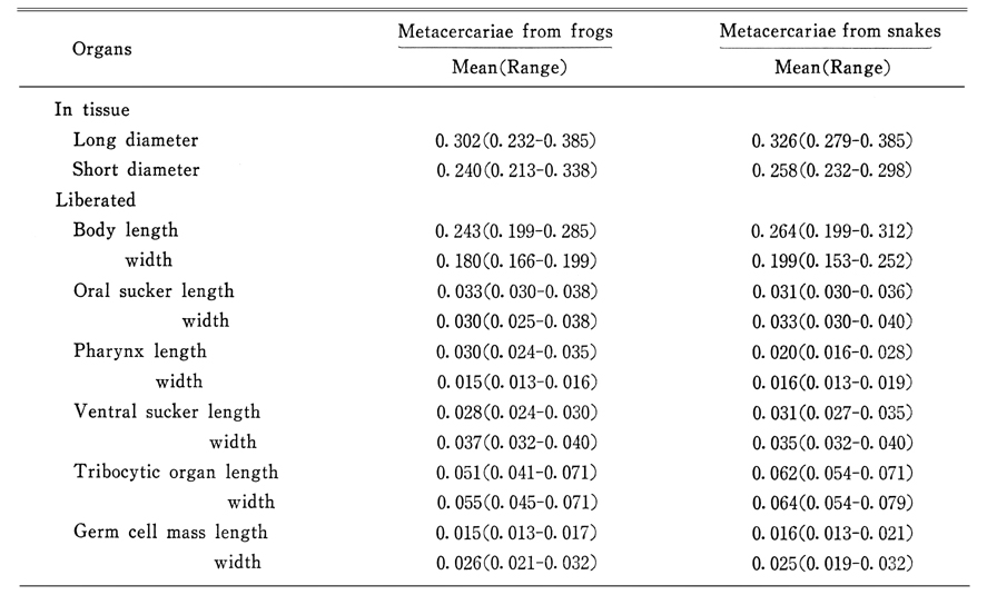
1. Rana nigromaculata and Natrix tigrina lateralis were found to be infected naturally by the metacercariae(diplostomula) of F. seoulensis. The metacercarial capsule was round to elliptical in tissue of the intermediate hosts with a long diameter 0.232~0.385 mm. Liberated metacercariae were ovoid with small conical posterior body. Body length measured 0.199~0.312 mm and width 0.153~0.252 mm.
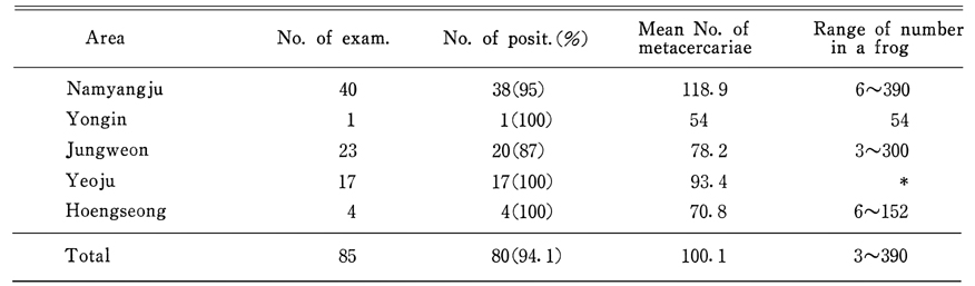
2. The infection rate of R. nigromaculata by the metacercariae ranged from 87% to 100% by area, and the number of the larvae ranged from 3 to 390 by frog. The metacercariae were found in skeletal muscle of frogs, from head to hindlegs.
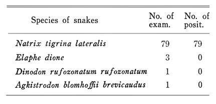
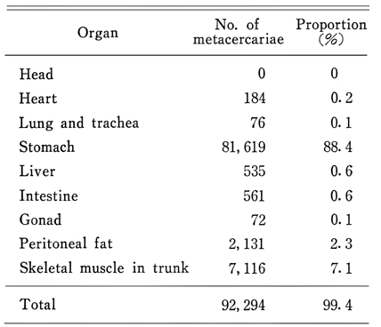
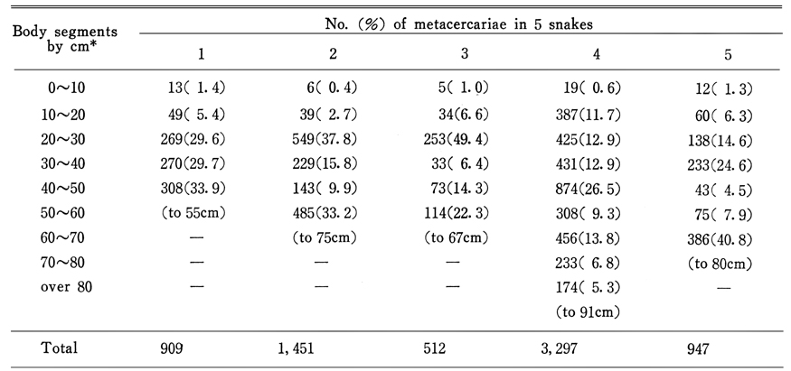
3. All examined N. t. lateralis were found to be infected by the metacercariae with the range of numbers frome 3 to 35,918. The larvae were collected from all viscera and body segments except for the head of the snakes. However, a great majority of the metacercariae were collected from the stomach.
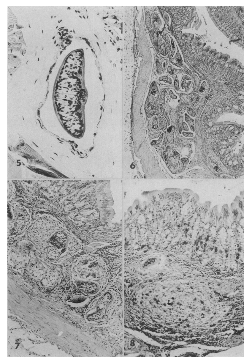
4. Hematoxylin-eosin stained preparations of frog skeletal muscle and snakes stomach revealed that the metacercariae had no cyst wall of worm origin, but encapsulated by the host tissue. Some of them were found in dilated lymphatic vessels. The larval infection was associated with slight or severe inflammatory reaction even with granuloma formation.
By above results, it was concluded that the frog, Rana nigromaculata, was the second intermediate host, and the snake Natrix tigrina lateralis was a paratenic host of F. seoulensis in nature in Korea.
Figures
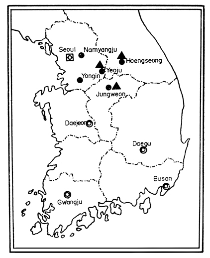
Fig. 1 The localities where the frogs(●) and snakes(▲) were collected.
EXPLANATIONS FOR PLATE Fig. 1. Macroscopic view of the metacercariae of F. seoulensis in snake omentum
Fig. 2. Low power view of the metacercaria of F. seoulensis in frog muscle, ×140
Fig. 3. The metacercaria of F. seoulensis obtained from a frog peritoneal cavity, unstained, ×280
Fig. 4. Liberated metacercaria of F. seoulensis from snake stomach, acetocarmine stained, ×280
EXPLANATIONS FOR PLATE Fig. 5. Sectioned metacercaria of F. seoulensis in endothelium lined duct in frog skeletal muscle, ×120
Fig. 6. Low power view of cross sectioned stomach of a snake showing many larvae of F. seoulensis in submucosa, HE stained, ×40
Fig. 7. Magnified view of submucosa showing tissue reactions around the metacercariae of F. seoulensis, ×100
Fig. 8. A granuloma with many eosinophils in submucosa of snake stomach, ×200
Tables
Table 1 Measurements of 10 acetocarmine stained metacercariae of F. seoulensis from frogs and snakes in millimeters
Table 2 Infection rate of frogs by the metacercariae of F. seoulensis
Table 3 Infection status of Rana nigromaculata by metacercariae of Fibricola seoulensis in the surveyed areas
Table 4 Distribution of metacercariae of Fibricola seoulensis by the body portion of frogs
Table 5 The infection of snakes by the metacercariae of F. seoulensis
Table 6 The number of metacercariae of F. seoulensis by the organ of Natrix tigrina lateralis*
Table 7 The number of the metacercariae of F. seoulensis from the body segments of N. t. lateralis
References
1.
Chandler AC. Trans Am Micr Soc 1942;61:156–167.
2.
Cook TW. J Parasit 1978;64:938–939.
3.
Hoffman GL. J Parasit 1955;41:327.
4.
Hong ST. Studies On Intestinal Trematodes In Korea: VII. Growth, Development And Recovery Of Fibricola Seoulensis From Experimentally Infected Rats And Mice. Korean J Parasitol 1982;20(2):112–121.
5.
Pearson JC. Observations on the morphology and life cycle of Neodiplostomum intermedium (trematoda: Diplostomatidae). Parasitology 1961;51:133–172.
6.
Seo BS, Cho SY, Hong ST, Hong SJ, Lee SH. Studies On Parasitic Helminths Of Korea 5.Survey On Intestinal Trematodes Of House Rats. Korean J Parasitol 1981;19(2):131–136.
7.
Seo BS, Lee SH, Hong ST, Hong SJ, Kim CY, Lee HY. Studies On Intestinal Trematodes In Korea: V. A Human Case Infected By Fibricola Seoulensis (Trematoda: Diplostomatidae). Korean J Parasitol 1982;20(2):93–99.
8.
Seo BS, Rim HJ, Lee CW. Studies on the parasitic helmiths of Korea: I. Trematodes of rodents. Korean J Parasitol 1964;2(1):20–26.
9.
Ulmer MJ. Notes on the morphology and host-parasite specificity of Fibricola cratera (Barker and Noll, 1915) Dubois 1932 (Trematoda: Diplostomatidae). J Parasitol 1955;41(5):460–466.