Warning: mkdir(): Permission denied in /home/virtual/lib/view_data.php on line 81

Warning: fopen(upload/ip_log/ip_log_2024-04.txt): failed to open stream: No such file or directory in /home/virtual/lib/view_data.php on line 83

Warning: fwrite() expects parameter 1 to be resource, boolean given in /home/virtual/lib/view_data.php on line 84
Studies on pathological changes of the liver in abino rats infected with Clonorchis sinensis
| Home | E-Submission | Sitemap | Contact us |  
top_img
Korean J Parasito Search

CLOSE

Korean J Parasito > Volume 16(2):1978 > Article

Original Article
Korean J Parasitol. 1978 Dec;16(2):148-155. English.
Published online Mar 20, 1994.  http://dx.doi.org/10.3347/kjp.1978.16.2.148
Copyright © 1978 by The Korean Society for Parasitology
Studies on pathological changes of the liver in abino rats infected with Clonorchis sinensis
Soon-Hyung Lee,Tae-Sub Shim,Sang Moon Lee and Je G. Chi
Department of Parasitology and Pediatrics, College of Medicine, Chung-Ang University, Korea.
Department of Pathology, College of Medicine, Seoul National University, Korea.
Abstract

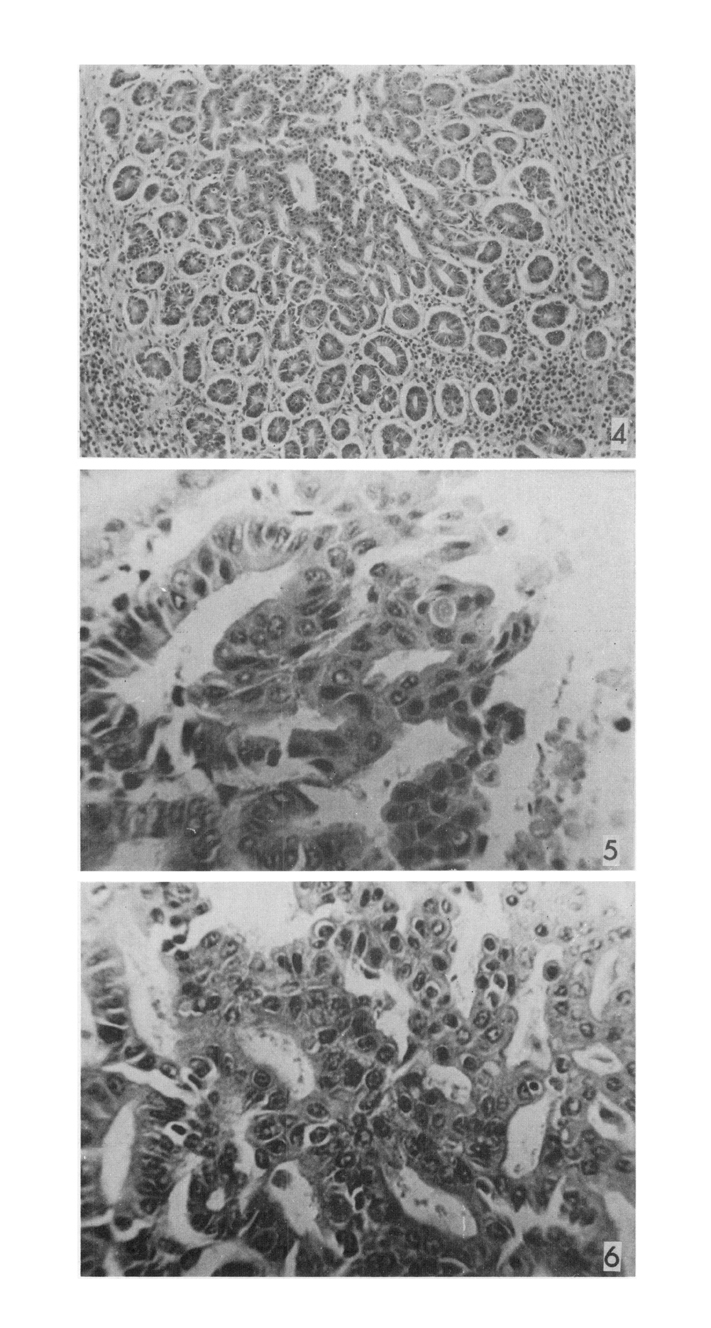
The present study was undertaken to observe the pathologic changes of the liver in albino rats with Clonorchis sinensis.

Twenty five rats given 50 metacercariae respectively by mouth were autopsied at 3 days (group 1), one week (group 2), 4 weeks (group 3), 8 weeks (group 4) and 12 weeks (group 5) of infection.

The following results were obtained:

1.Parasites were seen in bile ducts of group 2, 3, 4, 5 and increased in number with duration of infection.

2. The pathologic changes in the liver were prominent in intrahepatic bile ducts. Epithelial cells of bile ducts showed definite atypia and proliferation resulting in pseudostratification in group.

3. Stratification of metaplastic squamous cells and glandular proliferation were prominent in group 3. The epithelial cells were keratinized with syncitium and cribriform formation in group 4 and almost suggestive of adenomatous hyperplasia in group 5. Periductal fibrosis seen in group 4 was considerable as well as mature with hyalinized connective tissue in group 5.

4. Heavy inflammatory cell infiltrations around the affected bile ducts in group 1 became smaller in number with duration of infection. The inflammatory cells consisted of the majority of eosinophils in group 1 and chiefly plasma cells, lymphocytes and mononuclear cells in group 5.

5. Veins in portal spaces dilated markedly in group 1 became less prominent with duration of infection.

6. Although portal fibrosis increased definitely in group 3 often extended into the hepatic lobules in group 4, the changes of hepatic cells, sinusoids and central veins were negligible.

Above results suggest that clonorchiasis could be a factor in inducing primary carcinoma of liver in albino rats.

Figures


Figs. 1-3
Fig. 1. Glandular proliferation of the bile duct in C. sinensis infected albino rats. H & E stain, ×100.

Fig. 2. Stratification of metaplastic squamous epithelial cells of the bile duct. H & E stain, ×400.

Fig. 3. Downward proliferation of the lining epithelial cells froming many glandular structures. H & E stain, ×40.


Figs. 4-6
Fig. 4. High pover magnification of Fig. 3. Adenomatous proliferation of epithelial cells of bile duct. H & E stain, ×100.

Fig. 5. Keratinization of metaplastic squamous epithelial cells of bile duct. H & E stain, ×400.

Fig. 6. Cribriform pattern of metaplasitc squamous epithelial cells of bile duct. H & E stain, ×400.


References
1. Bae KW. Seoul J Med 1961;2(1):70–93.
2. Belamaric J. Intrahepatic bile duct carcinoma and C. sinensis infection in Hong Kong. Cancer 1973;31(2):468–473.
  
3. Ch'in KY, Lei AT, Wang TY. Primary mucinous carcinoma of liver associated with Clonorchis sinensis infection. Chin Med J 1955;73(1):26–35.
4. Chung KN. J Busan Med Coll 1975;15(1):217–227.
5. Chung CS, et al. Korean J Path 1976;10(1):33.
6. Faust EC, et al. Am J Hyg Mongr Ser 1927;8:1–207.
7. Germer WD, et al. Zeitschr Hyg Infe Ktionskrt 1955;141:132–139.
 
8. Hahn SS. Korean J Int Med 1961;4:351–365.
9. Hoeppli R. Chinese Med J 1933;47:1125–1141.
10. Hou PC. The pathology of Clonorchis sinensis infestation of the liver. J Pathol Bacteriol 1955;70(1):53–64.
  
11. Hou PC. The relationship between primary carcinoma of the liver and infestation with Clonorchis sinensis. J Pathol Bacteriol 1956;72(1):239–246.
  
12. Hou PC. Primary Carcinoma Of Bile Duct if The Liver if the Cat (Felis Catus) Infested With Clonorchis Sinensis. J Pathol Bacteriol 1964;87:239–244.
  
13. Chang HP. Pathological Changes in the Intrahepatic Bile Ducts of Cats (Felis Catus) Infested with Clonorchis Sinensis. J Pathol Bacteriol 1965;89:357–364.
  
14. Chang HP. Hepatic Clonorchiasis And Carcinoma Of The Bile Duct In A Dog. J Pathol Bacteriol 1965;89:365–367.
  
15. Kang TW. Seoul Univ J 1960;14:49–79.
16. Katsurada F. Beitrage zur pathologischen Anatomie und zur allgemeinen Pathologie 1900;28:479–505.
17. Kim JH. Korean J Int Med 1969;12(12):897–910.
18. Kim YI, et al. J Korean Surg Soc 1968;10(4):273–281.
19. Kim YI, et al. Seoul J Med 1974;15(3):247–253.
20. Lee CS. Theses Catholic Med Coll 1967;12:103–129.
21. Lee JK. J Cath Med Coll 1970;19:107–127.
22. Yoshimura H. Jap J Parasit 1972;21 Suppl:18–19.
23. Yoshimura H, et al. Jap J Parasit 1972;21(3):127–134.
Editorial Office
Department of Molecular Parasitology, Samsung Medical Center, School of Medicine, Sungkyunkwan University,
2066 Seobu-ro, Jangan-gu, Suwon 16419, Gyeonggi-do, Korea.
Tel: +82-31-299-6251   FAX: +82-1-299-6269   E-mail: kjp.editor@gmail.com
About |  Browse Articles |  Current Issue |  For Authors and Reviewers
Copyright © 2024 by The Korean Society for Parasitology and Tropical Medicine.     Developed in M2PI