Warning: mkdir(): Permission denied in /home/virtual/lib/view_data.php on line 81

Warning: fopen(upload/ip_log/ip_log_2024-05.txt): failed to open stream: No such file or directory in /home/virtual/lib/view_data.php on line 83

Warning: fwrite() expects parameter 1 to be resource, boolean given in /home/virtual/lib/view_data.php on line 84
An experimental study on the mouse intraperitoneal inoculation of the adult worm of Clonorchis sinensis
| Home | E-Submission | Sitemap | Contact us |  
top_img
Korean J Parasito Search

CLOSE

Korean J Parasito > Volume 15(2):1977 > Article

Original Article
Korean J Parasitol. 1977 Dec;15(2):75-92. English.
Published online Mar 20, 1994.  http://dx.doi.org/10.3347/kjp.1977.15.2.75
Copyright © 1977 by The Korean Society for Parasitology
An experimental study on the mouse intraperitoneal inoculation of the adult worm of Clonorchis sinensis
Chul Yong Song,Soon Hyung Lee and Je Geun Chi
Department of Parasitology, College of Medicine, Chung-Ang University, Korea.
Department of Pathology, College of Medicine, Seoul National University, Korea.
Abstract

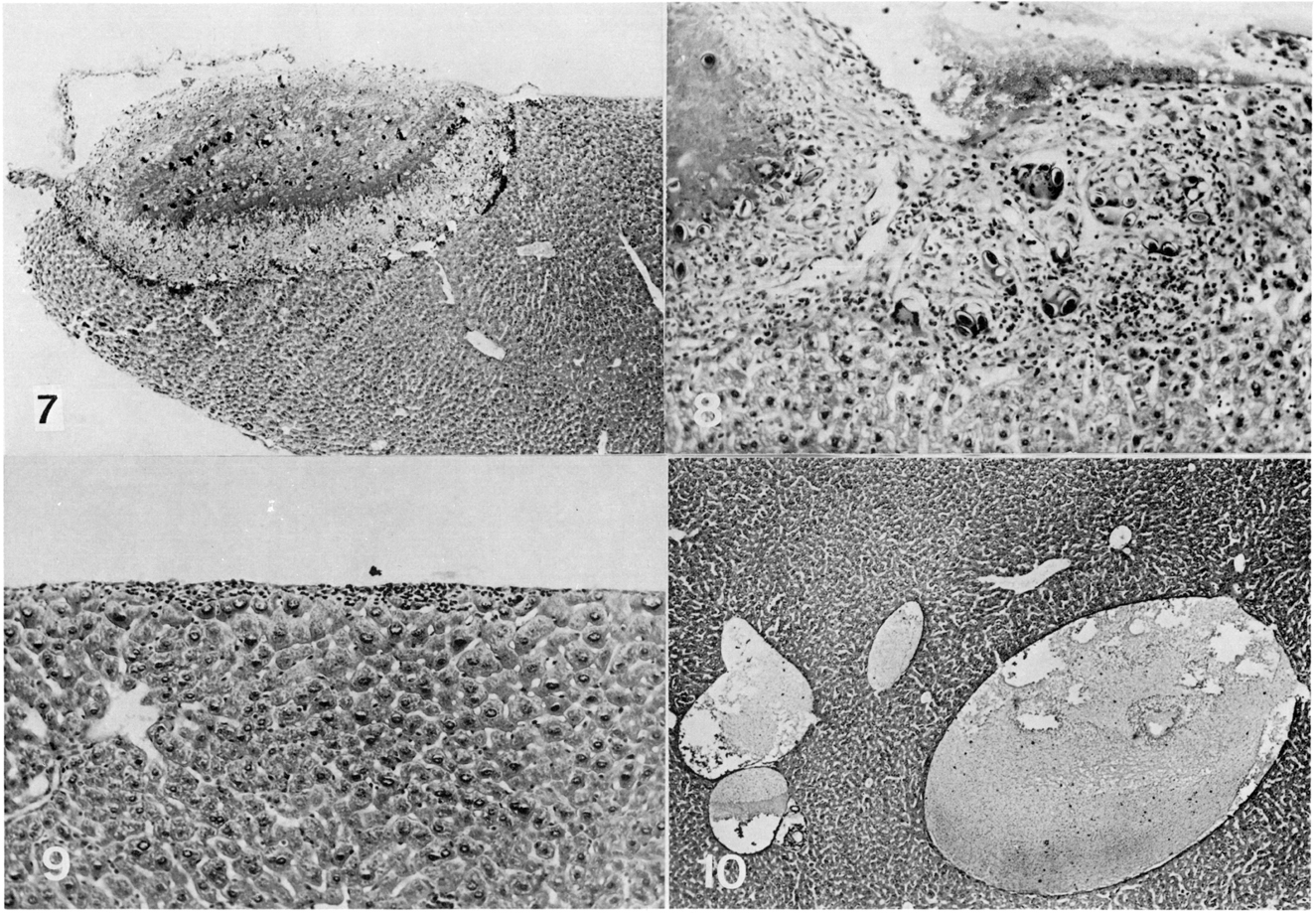
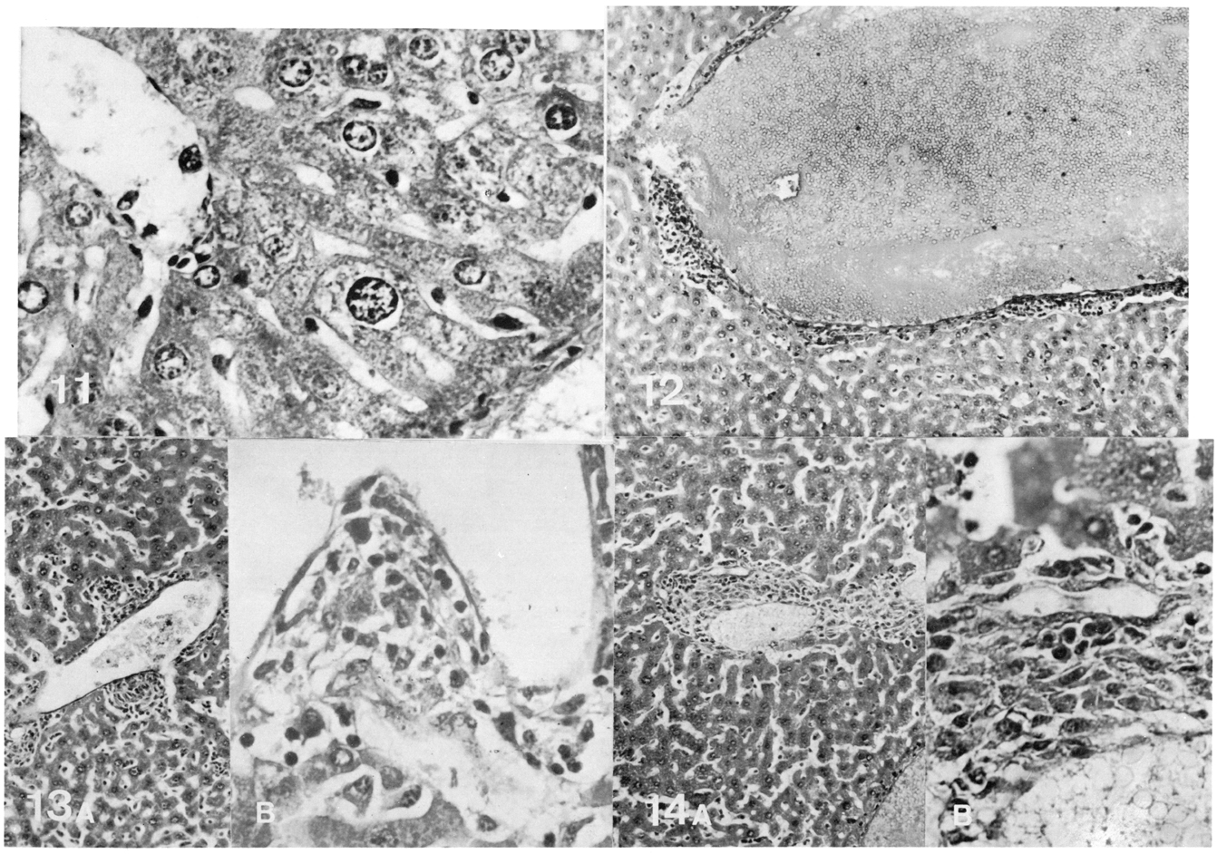
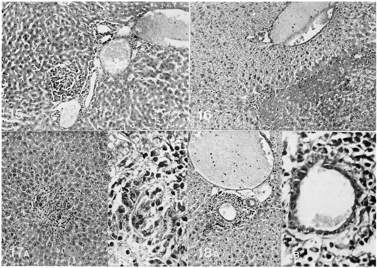
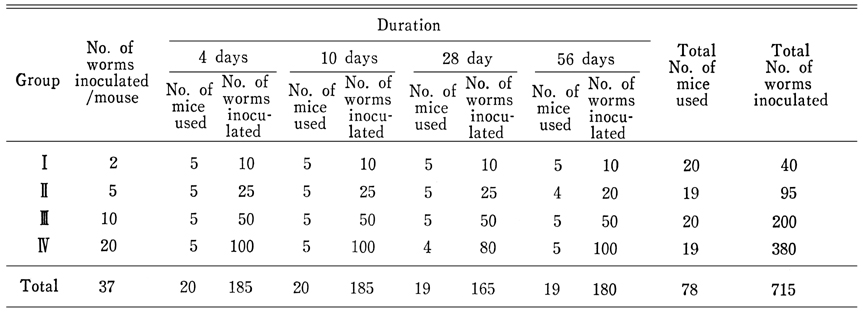
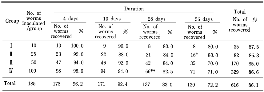
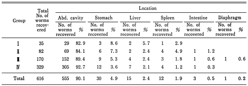
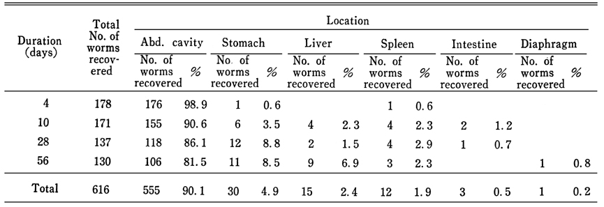
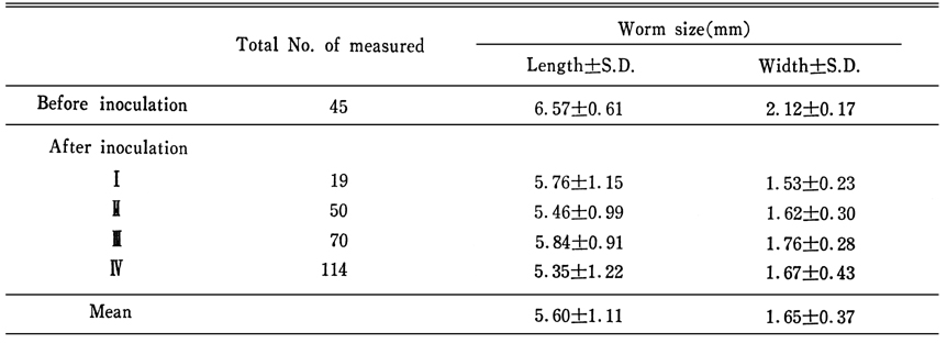
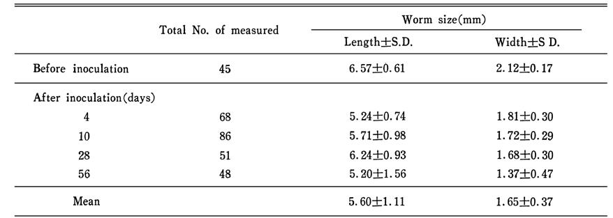
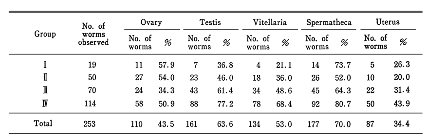
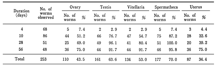
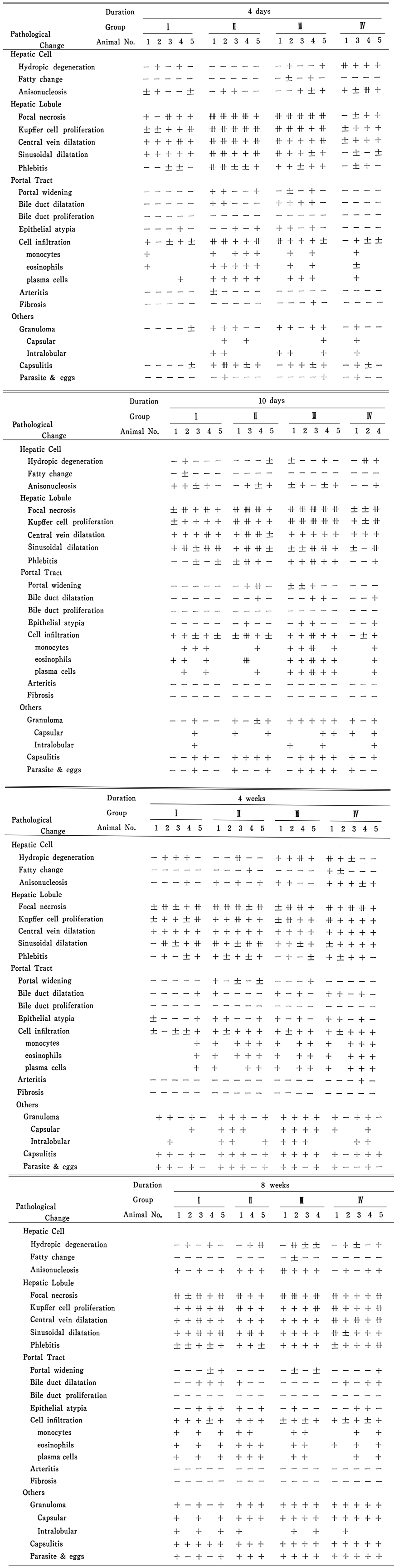
The present experimental study was undertaken to observe the chronological change of the worm structure of Clonorchis sinensis and the pathological findings of the liver when this fluke was inoculated to the mouse intraperitoneally.

The recovery rate, survival rate, location and size of the inoculated worms as well as the pathological changes of the liver were investigated for the comparison among the groups of mice, classified by number of worms and the duration of experiment.

The results obtained were summarized as follows.

1. The recovery and survival rates of the worms decreased especially 28 days after the inoculation.

2. Most of worms (90.l%) were collected from the peritoneal cavity and some of worms were found tightly adherent to the capsules of the liver, spleen, stomach, intestine and diaphragm. There were no worms recovered penetrated in the parenchymes of these organs.

3. The mean worm size after inoculation was smaller than that before inoculation. At the 10th day after the inoculation, the shrinkage of posterior portion of the worm body was observed.

4. Remarkable atrophy in the reproductive organs of the worm, such as spermatheca, testes, vitelline glands and ovary was frequently observed at the 10th day of inoculation.

5. Histopathologically the liver failed to show any parasitic worm inside the intrahepatic biliary system. However, multiple well formed egg-containing granulomas were present along the liver capsule. These necrotic granulomas were occasionally found under the fibrotic liver capsule. Focal necrosis and focal phlebitis together with vascular dilatation were prominent features seen in the liver. The bile duct in the liver showed mild dilation of the lumen, flattening of epithelial cells and periductal small round cell infiltration. Neither adenomatous hyperplasia nor portal fibrosis was seen in the whole experimental groups. Foci of intralobular micro-granulomas were found in some experimental animals.

6. The worms recovered in the capsule of the liver were degenerated and necrotized. Usually, there were remarkable capsulitis and granuloma formation around the eggs.

Figures


Figs. 1-6
Fig. 1. A normal worm (Clonorchis sinensis) of control group.

Fig. 2. A recovered worm from the abdominal cavity of the mouse, 4 days after the intraperitoneal inoculation. The spermatheca began to atrophy.

Fig. 3. This worm was obtained from the abdominal cavity 10 days after the inoculation. Note the shortened posterior portion of the worm.

Fig. 4. Atrophy of the spermatheca, tests, ovary, vitellaria and scantiness of eggs in the uterus were observed in the worm, 28 days after the inoculation.

Fig. 5. Remarkable atrophy of reproductive organs was visible in this worm, 28 days after the inoculation.

Fig. 6. Elongated or shortened worm bodies were obtained 56days after the inoculation. Note the atrophied reproductive organs.


Figs. 7-10
Fig. 7. Photomicrograph of a necrotic granuloma (presumed to me a dead worm) excavating the liver capsule. H&E ×40.

Fig. 8. Numerous Clonorchis eggs are surrounded by epitheloid granulomas. Langhans giant cell is seen. These granulomas are located just beneath the liver capsule. H&E ×40.

Fig. 9. The liver capsule showing a mild small round cell infiltration (capsulitis) along the surface. H&E ×100.

Fig. 10. Many dilated venous channels are seen. H&E ×40.


Figs. 11-14
Fig. 11. Hepatic lobule showing irregularity of nuclear size. H&E ×400.

Fig. 12. Dilated venous channel is seen in the center, the well of which is infiltrated with various inflammatory cells. The endothelial cells are also involved in the process. H&E ×100.

Fig. 13. Focal necrosis and cell collections including histiocytes are seen around a vein (A). Sometimes these cell collections protrude into the vascular lumen(B). H&E ×100(A) ×400(B).

Fig. 14. Perivascular cell infiltration consists mainly of lymphocytes and histiocytes. H&E ×100(A) ×400(B).


Figs. 15-18
Fig. 15. An area of focal necrosis in a lobule, showing small round cell infiltration alonf with liver cell degeneration. Histiocytes are lacking in this area. H&E ×100.

Fig. 16. Infarction of the liver is seen in the right lower portion.

Fig. 17. An intralobular granuloma with definite epitheloid cell component(A) and its magnification(B). H&E ×100(A) ×400(B).

Fig. 18. An intrahepatic bile duct exhibiting a mild luminal dilatation with eosinophilic secretion, epithelial flattening and a moderate periductal inflammatory cell infiltration. H&E ×100(A) ×400(B).


Tables


Table 1
Experimental design


Table 2
Number of worms recovered


Table 3
Number of worms survived


Table 4
Worms recovered from various parts of the body by group


Table 5
Worms recovered from various parts of the body by duration


Table 6
Worm size recovered before and after inoculation by group


Table 7
Worms size recovered before and after inoculation by duration


Table 8
Atrophic changes of reproductive organs of the worms found by group


Table 9
Atrophic changes of reproductive organs of the worms found by duration


Table 10
Histopathological changes of mouse liver

References
1. Chan PH, Teoh TB. The pathology of Clonorchis sinensis infestation of the pancreas. J Pathol Bacteriol 1967;93(1):185–189.
  
2. Chung CS, et al. Korean J Path 1976;10(1):33–46.
3. Chung KN. J Busan Med Coll 1975;15(1):217–227.
4. Faust EC, et al. Am J Hyg Monogr Ser 1927;8:1–207.
5. Hahn SS. Korean J Int Med 1961;4(6):417–444.
6. Hoeppli R. Chinese Med J 1933;47:1125–1141.
7. Hou PC. The pathology of Clonorchis sinensis infestation of the liver. J Pathol Bacteriol 1955;70(1):53–64.
  
8. Hou PC. The relationship between primary carcinoma of the liver and infestation with Clonorchis sinensis. J Pathol Bacteriol 1956;72(1):239–246.
  
9. Hou PC. Primary Carcinoma of Bile Duct of the Liver of the Cat (Felis Catus) Infested with Clonorchis Sinensis. J Pathol Bacteriol 1964;87:239–244.
  
10. Chang HP. Pathological Changes in the Intrahepatic Bile Ducts of Cats (Felis Catus) Infested with Clonorchis Sinensis. J Pathol Bacteriol 1965;89:357–364.
  
11. Chang HP. Hepatic Clonorchiasis and Carcinoma of The Bile Duct in A Dog. J Pathol Bacteriol 1965;89:365–367.
  
12. Kang TW. Seoul Univ J 1960;14:49–79.
13. Kim DH. Korean J Int Med 1966;9(1):33–48.
14. Kim JH. Korean J Int Med 1969;12(12):897–910.
15. Kim YI, et al. J Korean Surg Soc 1968;10(4):273–281.
16. Kim YI, et al. Seoul J Med 1974;15(3):247–253.
17. Kobayashi H. Saikingaku Zasshi 1912;202:1–66.
18. Komiya Y, et al. Jpn J Med Sci Biol 1953;6(6):571–575.
 
19. Lee CS. Theses Catholic Med Coll 1967;12:103–129.
20. Mukoyama T. J Jpn Path Soc 1921;11:443–446.
21. Rhee CH, Seo BS. [The Host-Parasite Relations Of Clonorchis Sinensis In The Mouse: Studies On The Development Of C. Sinensis In Mouse]. Korean J Parasitol 1968;6(3):101–109.
 
22. Seo BS. New Med J 1959;2(12):1389–1397.
23. Seo BS, et al. Seoul Univ Coll Theseon 1957;6:201–209.
24. Sun T, Chou ST, Gibson JB. Route of entry of Clonorchis sinensis to the mammalian liver. Exp Parasitol 1968;22(3):346–351.
  
25. Sun T, Gibson JB. Antigens of Clonorchis sinensis in experimental and human infections. Am J Trop Med Hyg 1969;18(2):241–252.
 
26. Yoshimura H. Jpn J Parasit 1965;14(5):445–451.
27. Yoshimura H. Jpn J Parasit 1972;21 Suppl:18–19.
28. Yoshimura H, et al. Jpn J Parasit 1972;21(3):127–134.
29. Wykoff DE. Studies on Clonorchis sinensis. III. The host-parasite relations in the rabbit and observations on the relative susceptibility of certain laboratory hosts. J Parasitol 1958;44(5):461–466.
  
30. Wykoff DE, Lepes TJ. Studies on Clonorchis sinensis. I. Observation on the route of migration in the definitive host. Am J Trop Med Hyg 1957;6(6):1061–1065.
 
Editorial Office
Department of Molecular Parasitology, Samsung Medical Center, School of Medicine, Sungkyunkwan University,
2066 Seobu-ro, Jangan-gu, Suwon 16419, Gyeonggi-do, Korea.
Tel: +82-31-299-6251   FAX: +82-1-299-6269   E-mail: kjp.editor@gmail.com
About |  Browse Articles |  Current Issue |  For Authors and Reviewers
Copyright © 2024 by The Korean Society for Parasitology and Tropical Medicine.     Developed in M2PI