Warning: mkdir(): Permission denied in /home/virtual/lib/view_data.php on line 81
Warning: fopen(upload/ip_log/ip_log_2024-04.txt): failed to open stream: No such file or directory in /home/virtual/lib/view_data.php on line 83
Warning: fwrite() expects parameter 1 to be resource, boolean given in /home/virtual/lib/view_data.php on line 84 Behaviour of mast cells in mice in the course of Entamoeba histolytica infection by strains
Behaviour of mast cells in mice in the course of Entamoeba histolytica infection by strains
Kyung-Il Im,Han-Ky Hwang and Chin-Thack Soh
Department of Parasitology, Yonsei University College of Medicine and Institute of Tropical Medicine, Yonsei University, Korea.
Abstract
The present report deals with the behaviour of mast cells in mice in the course of Entamoeba histolytica infection by the strains.
Mice weighing about l6 gm were used for three experimental groups; control, sham infection and experimental. The experimental group was infected with Entamoeba histolytica trophozoites directly into cecum by laparotomy. Strains isolated from three hepatic amoebic abscess cases were used. Mesenteric samples from the region of terminal ileum were fixed in methyl alcohol and stained with Pugh's solution. The ulcers in cecum were examined.
Changes in number and structure of mesenteric mast cells and blood eosinophils were as follows.
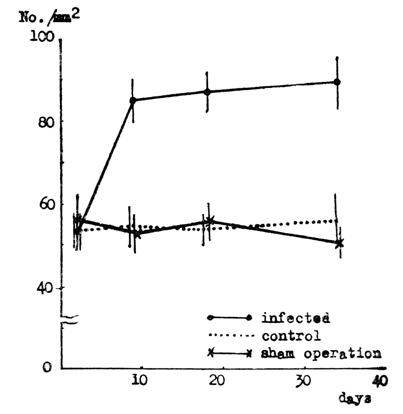
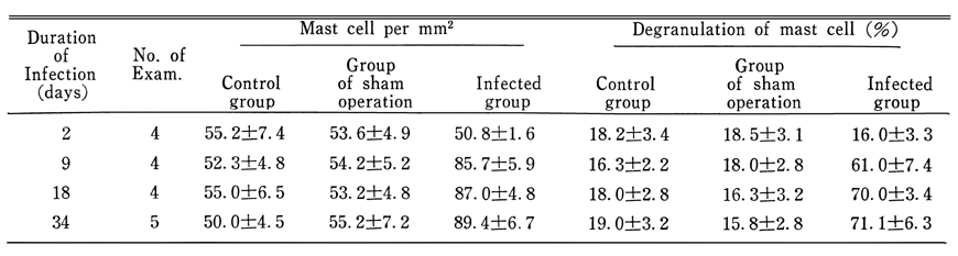
1. The number of mast cell in mesenteric tissues of the infected group increased from first day of the infection and persisted up to 34th day of the observation period.
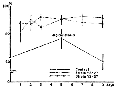
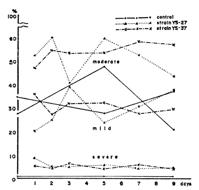
2. Degranulation and disruption of mast cells increased in the infected group compared with groups of the sham operation and the contro1, but showed no difference by the strains of Entamoeba histolytica.
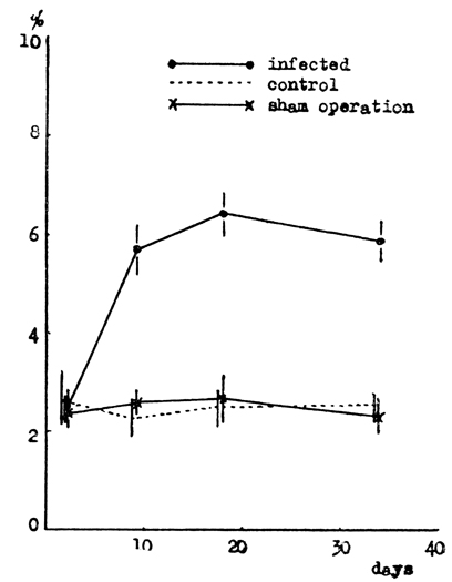
3. The blood eosinophilia was observed in the infected group and persisted until the observation period.
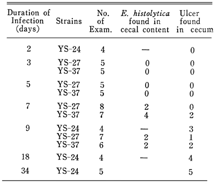
4. Ulcers in cecum were found in all the infected groups.
The results above indicate that mast cells are keenly related with the course of Entamoeba histolytica infection.
Figures
Fig. 1 Comparison of numbers of mesenteric mast cells per square millimeter in mice of each group.
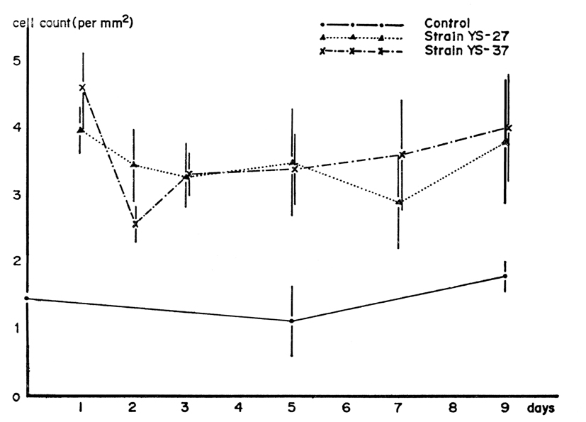
Fig. 2 Comparison of mast cell number in the mesentery of mice infected with Entamoeba histolytica by strains.
Fig. 3 Comparison of degranulation of mesenteric mast cells in mice infected with Entamoeba histolytica by strains.
Fig. 4 Comparison of degranulation status of mesenteric mast cells in mice infected with Entamoeba histolytica by strains.
Fig. 5 Comparison of percentage of blood eosinophils in each experimental group.
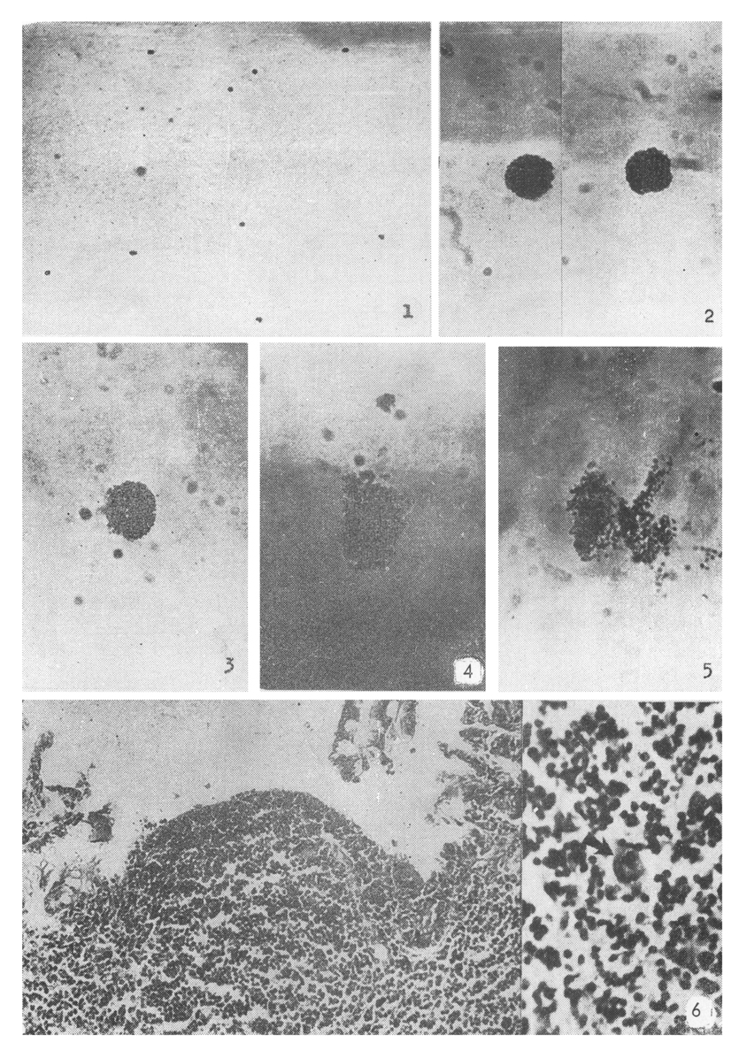
5. Disrupted mesenteric mast cell associated with marked, severe degranulation of metachromatic granules. (×450)
6. Ulcerated mucosa of the mouse cecum, and trophozoite of Entamoeba histolytica observed in this ulcer. (×450)
Tables
Table 1 Comparison of numbers of the mesenteric mast cells, and of degree of disrupted or degranulated mesenteric mast cells in mice infected with Entamoeba histolytica, strain YS-24
Table 2 Detection of Entamoeba histolytica in the cecal contents and ulcers in the infected cecum
References
1.
Blenkinsopp WK. Mast cell proliferation in adult mice. Nature 1967;214(5091):930–931.
2.
Bos HJ, et al. Z Parasitenk 1975;47:78–89.
3.
Campbell DH. J Infect Dis 1942;71:270–276.
4.
Carter PB, Higginbotham RD, Dougherty TF. The local response of tissue mast cells to antigen in sensitized mice. J Immunol 1957;79(3):259–264.
5.
Fernex M. Mast cells in the myocardium. Acta Trop 1961;18:177–187.
6.
Fernex M, Fernex P. Increased number of mast cells and helminthic diseases. Experimental mastocytosis in mice. Acta Trop 1962;19:248–251.
7.
Fernex M, Bezes H. Increased number of mast cells and helminthic diseases in man. Clinical observations. Acta Trop 1962;19:252–257.
8.
Goldgraber MB, Lewert RM. Immunological Injury of Mast Cells and Connective Tissue in Mice Infected with Strongyloides Ratti. J Parasitol 1965;51:169–174.
9.
Karmanska K, et al. Acta Parasit 1974;22:335–344.
10.
Lagunoff D, Benditt EP. Mast cell degranulation and histamine release observed in a new in vitro system. J Exp Med 1960;112:571–580.
11.
Lengy J, et al. J Parasit 1966;52:290.
12.
Riley JF. The effects of histamine-liberators on the mast cells of the rat. J Pathol Bacteriol 1953;65(2):471–479.
13.
Riley JF, West GB. The presence of histamine in tissue mast cells. J Physiol 1953;120(4):528–537.
14.
Smith DE. Nature of the secretory activity of the mast cell. Am J Physiol 1958;193(3):573–575.
15.
Speirs R, Wenck U, Dreisbach ME. Quantitative studies of the cellular responses to antigen injections in adrenalectomized mice. Blood 1956;11(1):56–70.
16.
Vaughn J. The stimulation of the eosinophil leucocyte. J Pathol Bacteriol 1952;64(1):91–102.
17.
Vaughn J. The function of the eosinophile leukocyte. Blood 1953;8(1):1–15.
18.
Wells PD. Mast cell, eosinophil and histamine levels in Nippostrongylus brasiliensis infected rats. Exp Parasitol 1962;12:82–101.
19.
West GB. Tissue mast cells and tissue amines. J Pharm Pharmacol 1959;11:513–534.