Warning: mkdir(): Permission denied in /home/virtual/lib/view_data.php on line 81

Warning: fopen(upload/ip_log/ip_log_2024-04.txt): failed to open stream: No such file or directory in /home/virtual/lib/view_data.php on line 83

Warning: fwrite() expects parameter 1 to be resource, boolean given in /home/virtual/lib/view_data.php on line 84
On the Sparganum mansoni infection in some Korean terrestrial snakes
| Home | E-Submission | Sitemap | Contact us |  
top_img
Korean J Parasito Search

CLOSE

Korean J Parasito > Volume 11(2):1973 > Article

Original Article
Korean J Parasitol. 1973 Aug;11(2):87-94. English.
Published online Mar 20, 1994.  http://dx.doi.org/10.3347/kjp.1973.11.2.87
Copyright © 1973 by The Korean Society for Parasitology
On the Sparganum mansoni infection in some Korean terrestrial snakes
Seung Yull Cho,Koo Il Hwang and Byong Seol Seo
Department of Parasitology and Institute of Endemic Diseases, College of Medicine, Seoul National University, Korea.
Abstract

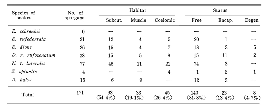
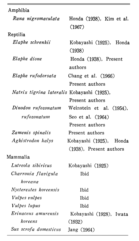
Distribution of Sparganum mansoni in 7 species of terrestrial snakes in Wonju City was surveyed. All kinds of snakes were found to be served as intermediate hosts of this larval worm except Elaphe schrenkii which has already been recorded as important host. Authors believe that Zamenis spinalis was firstly recorded as intermediate host of Sparganum mansoni in Korea.

Some aspects of infection status, host-parasite relations and sources of human infection were briefly discussed.

Figures


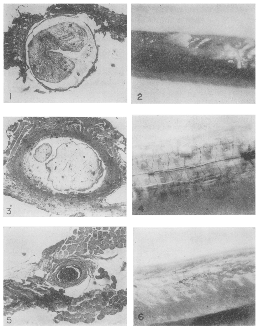
LEGENDS FOR FIGURES
Fig. 1: A section through mass of free stage of Sparganum mansoni (×30). Thin encapsulation with meager inflammatory response.

Fig. 2: The conglomerated masses of S. mansoni on the subcutaneous tissue of Agkistrodon halys.

Fig. 3: Section through encapsulated stage (×30). Viable worm is still present within wall and thick fibrous encapsulation is infiltrated by lymphocytes.

Fig. 4: Encapsulated stage of S. mansoni in subcutaneous tissue of Dinodon rufozonatum.

Fig. 5: Section through degenerative stage (×30). Amorphous eosinophilic material is filled within the granulomatous encapsulation.

Fig. 6: Elevated portion of snake muscle suggests the hidden intramuscular infection.


Tables


Table 1
Infection rate and worm burden of Sparganum mansoni in snakes


Table 2
Habitat and status of Sparganum in snakes


Table 3
The distribution of Sparganum in snakes by species


Table 4
Infectivity of three pathological stages of Sparganum to dogs


Table 5
List of animals which has been recorded as natural intermediate hosts of Sparganum mansoni in Korea since 1925

References
1. Chang K, et al. Theses Catholic Med Coll 1966;10:267–273.
2. Faust EC, et al. Am J Hyg 1929;9:560–583.
3. Honda D. J Chosen Med Ass 1938;28:1752.
4. Iwata S. Tokyo Med News 1932;2805:2784–2786.
5. Iwata S. Progress of Medical Parasitology in Japan 1962;2:279–321.
6. Jang DH. Korean J Vet Med 1964;8(4):3–11.
7. Kim CH, et al. Korean Modern Med 1967;6:191–195.
8. Kobayashi H. Jpn Med World 1925;5:9–16.
9. Kobayashi H. Keijo Med J 1928;9:109–124.
10. Mueller JF. J Parasitol 1937;23:308–310.
 
11. Seo BS, Rim HJ, Yoon JJ, Lee DJ. [A Case Report Of Sparganosis]. Korean J Parasitol 1964;2(3):179–182.
 
12. Weinstein PP, Krawczyk HJ, Peers JH. Sparganosis in Korea. Am J Trop Med Hyg 1954;3(1):112–129.
 
TOOLS
PDF Links  PDF Links
Full text via DOI  Full text via DOI
Download Citation  Download Citation
Share:      
METRICS
14
Crossref
0
Scopus
3,872
View
53
Download
A human case of Centrocestus armatus infection in Korea  1988 March;26(1)
Setaria marshalli infection in neonatal calves  1996 September;34(3)
Editorial Office
Department of Molecular Parasitology, Samsung Medical Center, School of Medicine, Sungkyunkwan University,
2066 Seobu-ro, Jangan-gu, Suwon 16419, Gyeonggi-do, Korea.
Tel: +82-31-299-6251   FAX: +82-1-299-6269   E-mail: kjp.editor@gmail.com
About |  Browse Articles |  Current Issue |  For Authors and Reviewers
Copyright © 2024 by The Korean Society for Parasitology and Tropical Medicine.     Developed in M2PI