Warning: mkdir(): Permission denied in /home/virtual/lib/view_data.php on line 81
Warning: fopen(upload/ip_log/ip_log_2024-04.txt): failed to open stream: No such file or directory in /home/virtual/lib/view_data.php on line 83
Warning: fwrite() expects parameter 1 to be resource, boolean given in /home/virtual/lib/view_data.php on line 84 Autoradiographic studies on the uptake and distribution of 14C-glucose by Paragonimus westermani
Autoradiographic studies on the uptake and distribution of 14C-glucose by Paragonimus westermani
Young Ok Park
Department of Parasitology, Institute of Endemic Diseases, College of Medicine, Seoul National University, Korea.
Abstract
Autoradiographic study was performed in order to know the distribution of exogenous 14C-glucose by lung fluke, Paragonimus westermani, incubated in Tyrode medium containing 10 uCi/ml of labeled substance.
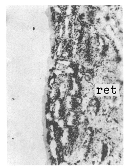
After 1 hour incubation at 37℃, microautoradiographs of this fluke showed that black grains derived from radioactive carbon were accumulated mainly in the parenchyme and subcuticular musculature. The muscular tissues such as oral sucker, pharynx and ventral sucker revaled considerable density of fine grains. Slight radioactivity was also observed in the regions of ovary, testes, vitelline follicles, eggs in uterus, intestinal ceca, and even in excretory bladder. Structures showing the least activity included the cuticle and uterine tubules of this fluke.
Figures
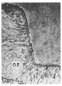
Fig. 1 Low density of fine grains were observed in oral sucker, mostly lined near the surface area (×100).
Fig. 2 Low density of fine grains were observed in oral sucker, mostly lined near the surface area (×100).
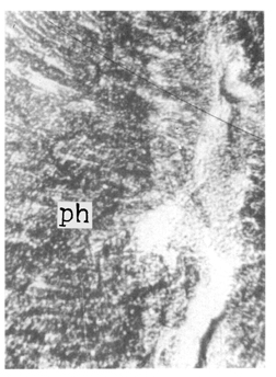
Fig. 3 The radioactivity was also detected in pharynx, higher than that of oral sucker (×100).
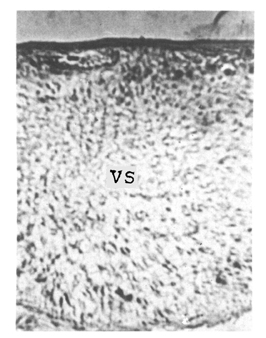
Fig. 4 Black grains were visible in the ventral sucker (×100). Relatively fine grains of them also accumulated near the outer surface.
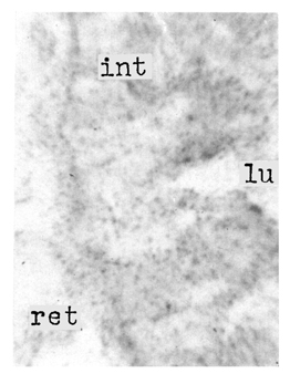
Fig. 5 High power magnification of intestinal wall revealed that grains were lined along the basement of the intestinal epithelium (×430).
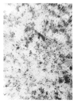
Fig. 6 Conspicuous black grains were observed inside of eggs, in uterus (×430).
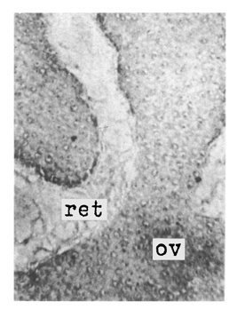
Fig. 7 In the region of ovary, slight radioactivity was monitored (×100).
Fig. 9 Microautoradiograhp of testes in which low density of grains were observed (×100).
Fig. 10 High power magnification of the testes (×430).
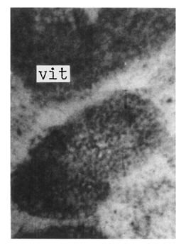
Fig. 11 High power magnification of vitelline follicles showed slight density of grains (×430).
Fig. 12 Some black grains lined along the wall of excretory bladder in high power magnification (×430).
References
1.
Burton PR. In vitro uptake of radioglucose by a frog lung-fluke and correlation with the histochemical identification of glycogen. J Parasitol 1962;48:874–882.
2.
Fitzgerald PJ, Simmel E, Weinstein J, Martin C. Radioautography; theory, technic, and applications. Lab Invest 1953;2(3):181–222.
3.
Hahn SS, et al. Korean J Int Med 1962;5:357–360.
4.
Halton DW. Studies on glycogen deposition in trematoda. Comp Biochem Physiol 1967;23(1):113–120.
5.
Kang IK, Lee SH, Seo BS. Study on the (14)C-glucose metabolism by Clonorchis sinensis: Paper Chromatographic Analyses in Combination with Autoradiography. Korean J Parasitol 1969;7(3):143–152.
6.
Lee EH, Seo BS. [Studies On Malic Dehydrogenase Activity In Parasitic Helminths]. Korean J Parasitol 1967;5(3):125–133.
7.
Lee SH. [Studies On Lactic Dehydrogenase Activity In Parasitic Helminths]. Korean J Parasitol 1967;5(1):5–16.
8.
Lotz WE, et al. Nucleonics 1953;11:54.
9.
Park CJ, Seo BS. [Studies On Phosphatase Activity In Some Parasitic Helminths]. Korean J Parasitol 1967;5(3):115–124.
10.
Tada I, et al. Jpn J Parasit 1961;10:490.
11.
Thorsell W, Appelgren LE, Kippar M. Distribution and fate of 2-C14-glucose in the liver fluke, Fasciola hepatica L., after short in vitro incubation. Z Parasitenkd 1968;31(2):113–121.