INTRODUCTION
Life largely depends on the dynamics of our climate system, especially the Earth’s surface climate. Although there has been much debate on whether or not climate change has any significant effect on the risk of vector-borne diseases and since the impact of climate change causing global warming on public health has been under consideration, its effect on vector-borne diseases has been at the forefront [1–3] and feasible changes in the prevalence of vector-borne diseases have attracted particular attention [4]. It is estimated that the worldwide average temperature will increase by 1.0–3.5°C by 2100 [5], thereby increasing the likelihood of many vector-borne diseases with public health significance by affecting the biology and ecology of their vectors and intermediate hosts and decisively increasing the risk of disease transmission [6,7] and consequentially, agricultural patterns, crop suitability and land use, and human communications. Thus, climate change as well as socio-economic status, vector control programs, drug resistance, etc. are likely to affect vector-borne disease epidemiology, from short-term epidemics to long-term gradual changes.
Tsutsugamushi disease, also known as scrub typhus, is caused by one of the world’s oldest recognized vector-borne pathogens, Orientia tsutsugamushi [8], which is a strictly intracellular gram-negative bacterium that requires host cells and is transmitted to humans by bites from the larvae of chigger mites. These arthropods serve as vectors of the disease whose endemicity is closely associated with their habitation [9–11]. It is a serious public health threat in the Asia-Pacific region and is the third most frequently reported infectious disease in Korea [12–15]. In endemic regions that have a high disease burden, the jeopardy of infection is mainly associated with agro-livestock and outdoor activities in rural regions [16]. The pivotal feature of the disease is being arthropod-mediated, maintaining the infection in nature through the relationship of warm-blooded animals including humans, mites, and the pathogen, O. tsutsugamushi. It causes disseminated vasculitic and perivascular inflammatory lesions resulting in significant vascular leakage and end-organ injury. After a latency period of 6–21 days, the disease onset is characterized by fever, headache, myalgia, cough, and gastrointestinal symptoms. A primary papular lesion which later crusts to form a flat black eschar can be present. If untreated, serious complications can occur involving various organs [17]. The endemicity of tsutsugamushi disease is closely related to the habitation of the chigger mite vector comprising 39 Leptotrombidium species carrying the intracellular pathogen O. tsutsugamushi found in Korea [18]. Leptotrombidium mites go through 4 stages: egg, larva (also called chigger), nymph, and adult. The larvae only feed on the body fluids of animal hosts, while nymphs and adults live by eating insect eggs under the leaves or in the ground [19]. At the present time, there is no vaccine against tsutsugamushi disease and general protective measures include avoiding exposure, wearing appropriate clothing, and using repellents to prevent chigger mite bites [12]. Basically, prevalence is one of the pivotal tools in the study of the epidemiology of chigger mites and chigger mite-borne parasites. Moreover, the role of chigger mites as vectors and the infected proportion are important tools for an adequate chigger mite control program, especially in terms of climate change.
In this study, we report on the species composition, species diversity, abundance, and distribution of chigger mites and their pathogens in Hwaseong-si, Gyeonggi-do (Hwaseong) in 2015 to monitor and reduce the potential for autochthonous transmission of chigger mite-borne Rickettsia spp. due to the effect of climate change in Korea. The results could provide the basis for future epidemiological studies and risk assessment of chigger mite-borne pathogens as an effect of climate change in Korea.
MATERIALS AND METHODS
Collection sites
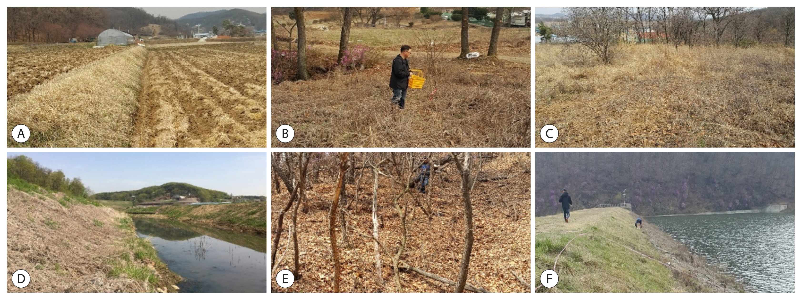
From April 2015 to November 2015, vector chigger mites potentially carrying tsutsugamushi disease (scrub typhus) collection was performed in Hwaseong from a rice field (Geographic Index (GI) recorded using a handheld global positioning system tracker: 37.183142/126.903973), vegetabe field (GI: 37.181500/126.899027), meadow (GI: 37.181218/126.900786), and waterway (GI: 37.183569/126.904895) 3 times (April, October, and November, 2015) with 5 collection traps at each site (Fig. 1). For the capture of rodents, 20 traps were set at each of the collection sites: the rice field (GI: 37.182406/126.903726), vegetable field (GI: 37.181603/126.900196), waterway (GI: 37.189779/126.902948), meadow (GI: 37.182490/126.904874), and hill (GI: 37.181458/126.898072) in April, October, and November 2015.
Collecting chigger mites and rodents
Chigger mites were collected to ensure their presence and for O. tsutsugamushi detection in Hwaseong using chigger mite collecting traps mimicking human skin odor and sticky chigger traps (size: 250×187 mm (H) with a sticky board: 20×200 ×0.8 mm (T)) on the surface using octenol and lactic acid as a lure. A total of 100 traps were set at 5 points, as described earlier. Adhesive tape on the surface of the mite traps was collected biweekly. The chigger mite samples were tested for O. tsutsugamushi and all of the mites were classified morphologically. The information regarding all of the collected specimens including their location, the number of mites collected from the body of each host animal, and the date of collection were recorded. The chigger mites were removed from the tape under a microscope, placed in 80% ethanol, mounted on glass slides with mounting media, and then their species identified using standard key morphological features for chigger mites in Korea [20].
A total of 100 Sherman collapsible rodent traps were set each night in a variety of habitats (20 traps per site) (Fig. 2). They were placed in a grid formation every 10 m according to the land topology and were baited with appropriate food. The collected rodents were transported to the laboratory alive and their species identified using taxonomic keys [20]. Following transfer to the laboratory, cervical dislocation was used to euthanize the transferred rodents, after which the hosts’ information was recorded and they were hung upside down for 48 hr to gather the chigger mites; Petri dishes with distilled water were installed under the rodents to prevent drying. After 48 hr, laboratory staff recorded and gathered the chigger mites falling from the rodents using sterile needles under an inverted microscope. The total number of chigger mites recovered from the rodent hosts was counted: half of the collected chigger mite samples were tested for O. tsutsugamushi and the rest were classified morphologically.
Preparation of specimens for species identification
Chigger mites were individually isolated from the tape of the sticky chigger mite traps using sterile needles and kerosene under an inverted microscope. Number of chigger mites recovered from the tape was counted. They, 109 mites, were carefully recovered, washed with absolute ethanol, and mounted with PVA MTNG medium on glass slides, after which they were dried at room temperature for 24 hr. In the case of the chigger mites recovered from the captured rodents, a representative number (approximately 50%) was sampled due to the practical difficulties of examining all 4,139 chigger mites. Glass-slide-mounted specimens were examined under a dissecting microscope at ×400 magnification. They were identified after recording their general characteristics, including body shape and color variation; standard taxonomic keys described by Ree [20] were used to identify the genera. The chigger mites were then pooled (1–50/pool) according to genus and rodent host.
Detection and identification of pathogens
A one-step real-time RT-qPCR assay for the detection of pathogens in chigger mites was conducted using a Verso SYBR Green 1-step RT-qPCR kit (Thermo AB4104, Thermo-Fisher Scientific, Waltham, Massachusetts, USA) in accordance with the manufacturer’s protocol.
The pathogen O. tsutsugamushi for tsutsugamushi disease was detected using an INNOPLEXTM TSUTSU detection kit (iNtRON Biotechnology, Seoul, Korea) with DNA samples prepared using a G-spinTM total DNA extraction kit (iNtRON Biotechnology) via the nested-PCR method in accordance with the manufacturer’s protocol. The species-specific primers used for protein antigen gene 56 kDa from O. tsutsugamushi were as follows: WJ173 (OutForward) 5′-CCAGGATTTAG-AGCAGAG-3′, and WJ174 (OutReverse) 5′-CGCTAGGTTTATTAGCAT-3′ (amplified DNA band size: 509 bp), and WJ175 (InForward) 5′-CCTCAGCCTACTATRAKKCC-3′ and WJ176 (InReverse) 5′-AGCATTTGATAATGCAGCAAGACC-3′ (amplified DNA band size 350 bp). After performing nested-PCR and electrophoresis on a 1% agarose gel, the identified DNA bands were used for the pathogen detection by means of DNA sequencing. The analysis of the derived nucleotide sequences was performed for matching genotypes using the NCBI-BLAST service.
RESULTS
Chigger mites collectied
A total of 109 chigger mites were collected in April, October, and November from Hwaseong (Tables 1, 2). Five species were identified and distributed among 4 genera: L. scutellare (n =66/60.6%), L. pallidum (n=31/28.4%), Neotrombicula tamiyai (n=10/9.2%), Euschoengastia koreaensis (n=1/0.9%), and Neoschoengastia asakawa (n=1/0.9%). Leptotrombidium was the most frequently collected mite genus (89.0%), most often from the rice field sites (Table 1). Of the 3 months, the highest collection of chigger mites using sticky chigger traps was during October 2015 (the autumn season), while no mites were collected in April (Table 2). However, in the waterway, the highest collection level was during November.
Apodemus agrarius in Hwaseong area
A total of 77 small rodents were captured during 6 trapping nights (Table 3). Table S1 gives the rodents captured and species composition at the trapping times. Rodent species composition was found to be relatively similar at all of the trapping surveillance sites and times. From the 77 rodents examined, all were found to be infected with ectoparasites; nearly all had chigger mites (89.6%, 69/77, Supplementary Table S1), with a mean abundance (chigger index) of 53.8. Rodents with chigger mite infection were found at all trapping areas surveyed. The highest number of chigger mites in the H-10-2 rodent was 432 and was obtained from a trap during the second survey in April 2015, which accounted for 10.4% of the total recovery. Of the 4,139 chigger mites, 96.5% were from Apodemus agrarius, while 3.5% were from Micromys minutus. Less than 1% were recovered from Crocidura lasiura, suggesting that A. agrarius is the main rodent host in the surveyed Hwaseong areas (Supplementary Table S1). Nine species were identified and distributed in 3 genera: Leptotrombidium (n=1,959/94.8%), Euschoengastia (n=72/3.5%), and Neotrombicula (n=34/1.6%). Of the 2,065 chigger mites examined, 46.9% (n=969) were L. pallidum, while 18.6% (n=384) were L. scutellare, 18.0% (n=372) were L. orientale, 7.7% (n=159) were L. zetum, 3.5% (n=73) were L. palpale, 3.5% (n=72) were Eu. koreaensis, and 1.4% (n=28) were N. tamiyai (Table 4). The remaining 2 species comprised less than 1% of the total number of chigger mites collected in this study. Leptotrombidium was the most frequently collected mite genus (1,959/2,065, 94.9%). Nearly all rodents with ectoparasites had polyspecific parasitism comprising mites, Laelaptidae, ticks, fleas, lice, and others (Supplementary Table S1). In 77 wild rodents, 2,074 of the total of 4,139 chigger mites (approximately 50%) were used as specimens for separating the pathogens for tsutsugamushi disease. Of the chigger mites screened using an INNOPLEXTM TSUTSU detection kit (iNtRON Biotechnology) with DNA samples by the nested-PCR method, none of the pools recovered from the rodent hosts tested positive for O. tsutsugamushi: O. tsutsugamushi primers failed to amplify the chigger mite samples, although they did amplify the O. tsutsugamushi positive control. The rest of the collected chigger mites (2,065) were used to determine the scientific names of the identified species (Table 3).
DISCUSSION
Many recent studies from various regions provide evidence to indicate that infectious diseases such as flaviviral diseases and tsutsugamushi disease are no longer restricted to their first-identified habitats and that the global resurgence of vector-borne infectious diseases and their potential has emerged as a critical public health issue, especially owing to climate change. Serological and molecular evidence of infectious diseases has been accumulating.
In this study, we surveyed the species composition, species diversity, abundance, and distribution of chigger mites and their pathogens in Hwaseong in 2015. In Korea, it is generally accepted that L. pallidum and L. scutellare are the predominant vector species and are distributed throughout the Korean Peninsula [18,21]. Park and Shin [22] determined that L. pallidum is the predominant species (74.9%), followed by L. scutellare (18.9%) and L. palpale (2.7%), and that L. pallidum and L. scutellare are the most prevalent scrub typhus vectors. In addition, Roh et al. [21] reported that, in Korea, the predominant mite species were L. pallidum, (52.6%), L. scutellare (27.1%), L. palpale (8.2%), L. orientale (5.6%), and N. tamiyai (1.7%) and that the geographical distribution map of the L. scutellare chigger mite index (total number of chigger mites divided by total number of used traps) was identical to the incidence pattern of tsutsugamushi disease. From another report by Park and Shin [22], the predominant chigger mite species collected during the spring and fall seasons from A. agrarius were L. pallidum (57.6%), L. palpale (14.5%) and L. scutellare (7.9%). However, L. scutellare was collected only along the southeastern coast in Yeongduk-gun (county) area, Gyeongsangbuk-do. Our findings from the captured rodents are similar to previous reports in that L. scutellare was the predominant chigger mite species (60.6% rather than 28.4% for L. pallidum) from the collection of the hosts with sticky chigger traps [21–23].
In general, the mite species known to be important as the potential vectors of O. tsutsugamushi are widely distributed throughout the world [24]. In addition, it is well known that chigger mite development is temperature-dependent. From 2010 to 2013, a total of 27,791 cases of tsutsugamushi disease throughout the nation were reported to the KCDC [25] and the incidence of tsutsugamushi disease has increased steadily [13,26]. Recently, the incidence of tsutsugamushi disease has expanded upwardly to the Northern region due to the contribution of global warming [13,22,27]. In addition, although it is thought to be a rural disease, indigenous urban cases have been investigated in cities, especially the metropolitan cities of Korea [13]. This study provides pivotal scientific data that identifies potential tsutsugamushi disease risks for the development of preventive and control measures against it in Gyeonggi-do. However, future and continuous surveillance should be aimed at the rodent host sample size and diversity and greater detail of the overall geographic distribution of chigger mites.
In this study, we tried to determine the distribution of tsutsugamushi disease (scrub typhus) transmitted by chigger mites living on rodents and to investigate the target vector diversity, abundance, and distribution to enable the mapping of hotspots for this disease. Along with the impact of climate change, the distribution of vectors and reservoir hosts are pivotal aspects of understanding the epidemiology of the diseases and the risk to public health. Further studies are needed to remain vigilant by monitoring the vectors of suspected vector-borne diseases to reduce the potential for transmission to resident populations. Given increasing global networking and global warming, the risk for invasion of the vector-borne diseases via active human life and transportation has become continual, and so enhanced monitoring and long-term surveillance of them are of great public health interest.
Supplementary Information
Supplementary Table S1
The total number and identity of rodent hosts and ectoparasites collected in Hwaseong for 2 days of each month in 2015
| Collection date | Host identity | Host | Ectoparasite | |||||||||||||
|---|---|---|---|---|---|---|---|---|---|---|---|---|---|---|---|---|
|
|
|
|||||||||||||||
| Species* | Sex | Wt (g) | Head+ body (mm)+ | Tail (mm) | Total size (mm) | Ear (mm) | Right foot (mm) | Mite | Laelaptidae | Tick | Flea | Lice | Others | |||
| April | 1st | H-4-1 | M. minutus | F | 15 | 8.7 | 7.5 | 12.1 | 1.1 | 0.9 | 143 | 0 | 0 | 0 | 0 | 294 |
| H-4-2 | A. agrarius | M | 40 | 11.2 | 9.3 | 20.5 | 1.5 | 1.7 | 53 | 5 | 0 | 0 | 69 | 44 | ||
| H-4-3 | A. agrarius | F | 28 | 9.9 | 6.2 | 16.1 | 1.5 | 1.7 | 68 | 10 | 0 | 0 | 4 | 3 | ||
| H-4-4 | A. agrarius | M | 35 | 10.1 | 10.4 | 20.5 | 1.4 | 1.6 | 196 | 9 | 0 | 1 | 14 | 7 | ||
| H-4-5 | A. agrarius | F | 23 | 9.1 | 8.3 | 17.4 | 1.2 | 1.4 | 90 | 1 | 0 | 0 | 2 | 0 | ||
| H-4-6 | A. agrarius | M | 26 | 10.2 | 7.7 | 17.9 | 1.3 | 1.4 | 41 | 0 | 0 | 0 | 24 | 5 | ||
| H-4-7 | A. agrarius | M | 36 | 11 | 9 | 20 | 1.5 | 1.7 | 67 | 3 | 1 | 0 | 4 | 0 | ||
| H-4-8 | A. agrarius | F | 40 | 10 | 8.5 | 18.5 | 1.3 | 1.6 | 38 | 4 | 1 | 1 | 3 | 2 | ||
| H-4-9 | A. agrarius | M | 25 | 9.8 | 8.3 | 18.1 | 1.4 | 1.4 | 161 | 4 | 1 | 0 | 3 | 0 | ||
| H-4-10 | A. agrarius | F | 24 | 9.4 | 8.5 | 17.9 | 1.4 | 1.4 | 148 | 5 | 1 | 0 | 1 | 0 | ||
| H-4-11 | A. agrarius | F | 28 | 9.8 | 8.2 | 18 | 1.3 | 1.5 | 53 | 3 | 1 | 1 | 2 | 0 | ||
| H-4-12 | A. agrarius | M | 20 | 9.5 | 7.5 | 18 | 1.3 | 1.5 | 122 | 0 | 0 | 1 | 2 | 0 | ||
| H-4-13 | A. agrarius | F | 25 | 10.5 | 8.1 | 18.6 | 1.6 | 1.5 | 17 | 1 | 0 | 1 | 498 | 0 | ||
| H-4-14 | C. lasiura | F | 14 | 7.1 | 4.2 | 11.3 | 0.9 | 1 | 0 | 0 | 1 | 0 | 0 | 0 | ||
| H-4-15 | C. lasiura | F | 15 | 7.5 | 4.5 | 12 | 1.1 | 1 | 0 | 0 | 0 | 1 | 29 | 0 | ||
| H-4-16 | A. agrarius | F | 32 | 11 | 8.4 | 19.4 | 1.5 | 1.7 | 27 | 0 | 0 | 0 | 0 | 1 | ||
| H-4-17 | A. agrarius | F | 25 | 10.2 | 7.7 | 17.7 | 1.4 | 1.5 | 3 | 1 | 0 | 2 | 1 | 0 | ||
| H-4-18 | A. agrarius | F | 25 | 10.1 | 8.7 | 18.8 | 1.3 | 1.5 | 74 | 0 | 0 | 0 | 11 | 1 | ||
| H-4-19 | A. agrarius | F | 22 | 9.2 | 8.2 | 17.4 | 1.4 | 1.5 | 0 | 2 | 0 | 0 | 11 | 0 | ||
| H-4-20 | A. agrarius | M | 30 | 10.8 | 6.1 | 16.9 | 1.4 | 1.4 | 9 | 0 | 0 | 2 | 5 | 0 | ||
| 2nd | H-4-21 | A. agrarius | F | 30 | 11.2 | 9.2 | 20.4 | 1.4 | 1.5 | 7 | 1 | 0 | 1 | 1 | 2 | |
| H-4-22 | A. agrarius | M | 35 | 11.2 | 9.2 | 20.4 | 1.4 | 1.5 | 20 | 2 | 0 | 0 | 1 | 0 | ||
| H-4-23 | A. agrarius | F | 10 | 6.5 | 5.5 | 12 | 0.7 | 1.2 | 1 | 3 | 0 | 0 | 1 | 0 | ||
| H-4-24 | A. agrarius | F | 30 | 11 | 7.8 | 18.8 | 1.4 | 1.4 | 18 | 0 | 0 | 0 | 0 | 4 | ||
| H-4-25 | A. agrarius | F | 30 | 10.5 | 8 | 18.5 | 1.4 | 1.4 | 57 | 0 | 0 | 0 | 1 | 11 | ||
| H-4-26 | C. lasiura | M | 8 | 8 | 3.8 | 11.8 | 0.6 | 1.1 | 1 | 0 | 0 | 0 | 0 | 0 | ||
| H-4-27 | A. agrarius | M | 28 | 28 | 8.5 | 18.9 | 1.4 | 1.5 | 16 | 0 | 0 | 0 | 0 | 0 | ||
| H-4-28 | A. agrarius | F | 33 | 33 | 8 | 19 | 1.5 | 1.6 | 18 | 0 | 0 | 0 | 0 | 2 | ||
| H-4-29 | A. agrarius | F | 30 | 30 | 8.5 | 19.7 | 1.6 | 1.6 | 0 | 1 | 0 | 3 | 3 | 0 | ||
| H-4-30 | A. agrarius | M | 50 | 50 | 9.5 | 22 | 1.5 | 1.7 | 5 | 0 | 0 | 1 | 2 | 3 | ||
|
|
||||||||||||||||
| October | 1st | H-10-1 | A. agrarius | M | 34 | 8.7 | 9.5 | 18.2 | 1.4 | 1.4 | 68 | 2 | 0 | 0 | 0 | 0 |
| H-10-2 | A. agrarius | M | 40 | 13 | 8 | 21 | 1.4 | 1.2 | 432 | 2 | 0 | 0 | 1 | 0 | ||
| H-10-3 | A. agrarius | F | 32 | 9 | 9.6 | 18.6 | 1.2 | 1.4 | 194 | 2 | 0 | 0 | 0 | 0 | ||
| H-10-4 | A. agrarius | M | 40 | 9.7 | 10.4 | 20.1 | 1 | 1.2 | 28 | 1 | 0 | 0 | 2 | 0 | ||
| H-10-5 | A. agrarius | F | 33 | 8.5 | 9.2 | 17.7 | 1.2 | 1.1 | 155 | 0 | 0 | 0 | 0 | 0 | ||
| H-10-6 | A. agrarius | F | 26 | 8.3 | 8.2 | 16.5 | 1.5 | 1.3 | 12 | 1 | 1 | 0 | 0 | 0 | ||
| H-10-7 | A. agrarius | F | 17 | 7 | 7.5 | 14.5 | 1.3 | 1.3 | 37 | 5 | 0 | 0 | 0 | 0 | ||
| 2nd | H-10-8 | A. agrarius | F | 51 | 11.5 | 10 | 21.5 | 1.2 | 1.5 | 59 | 7 | 0 | 0 | 0 | 0 | |
| H-10-9 | A. agrarius | M | 44 | 13 | 5.8 | 18.8 | 1.2 | 1.4 | 27 | 1 | 0 | 0 | 1 | 0 | ||
| H-10-10 | A. agrarius | M | 47 | 13.5 | 9.5 | 23 | 1 | 1.4 | 18 | 0 | 0 | 0 | 0 | 0 | ||
| H-10-11 | A. agrarius | M | 35 | 10.3 | 8 | 18.3 | 1.2 | 1.3 | 4 | 0 | 0 | 0 | 0 | 0 | ||
| H-10-12 | A. agrarius | F | 37 | 11.7 | 10.5 | 22.2 | 1.1 | 1.4 | 18 | 11 | 1 | 0 | 0 | 0 | ||
| H-10-13 | A. agrarius | M | 42 | 12.1 | 11 | 23.1 | 1.3 | 1.3 | 7 | 2 | 0 | 0 | 0 | 0 | ||
| H-10-14 | A. agrarius | F | 34 | 11.8 | 9 | 20.8 | 1.2 | 1.3 | 18 | 0 | 1 | 0 | 0 | 0 | ||
| H-10-15 | A. agrarius | M | 21 | 8 | 6 | 14 | 0.9 | 1 | 131 | 22 | 1 | 0 | 10 | 0 | ||
| H-10-16 | A. agrarius | M | 20 | 8 | 6.3 | 14.3 | 1.1 | 1.2 | 2 | 2 | 3 | 0 | 4 | 0 | ||
| H-10-17 | A. agrarius | F | 29 | 12.5 | 10 | 22.5 | 1.3 | 1.4 | 1 | 1 | 0 | 0 | 4 | 0 | ||
| H-10-18 | C. lasiura | M | 12 | 7 | 4.3 | 11.3 | 0.5 | 0.5 | 0 | 2 | 3 | 0 | 0 | 0 | ||
|
|
||||||||||||||||
| November | 1st | H-11-1 | A. agrarius | M | 17 | 9 | 8.5 | 17.5 | 1 | 1.4 | 391 | 5 | 2 | 3 | 0 | 0 |
| H-11-2 | A. agrarius | M | 32 | 11 | 10 | 21 | 1.2 | 1.3 | 26 | 5 | 0 | 0 | 0 | 0 | ||
| H-11-3 | A. agrarius | M | 24 | 11 | 8 | 19 | 1.2 | 1.5 | 60 | 8 | 0 | 0 | 0 | 1 | ||
| H-11-4 | A. agrarius | F | 38 | 13 | 8 | 21 | 1.2 | 1.3 | 13 | 0 | 0 | 0 | 3 | 0 | ||
| H-11-5 | A. agrarius | M | 24 | 10 | 8 | 18 | 1 | 1.4 | 22 | 4 | 0 | 0 | 0 | 7 | ||
| H-11-6 | A. agrarius | F | 29 | 12 | 9 | 21 | 1.2 | 1.4 | 46 | 3 | 0 | 0 | 0 | 0 | ||
| H-11-7 | A. agrarius | M | 18 | 10.5 | 6.5 | 17 | 0.9 | 1 | 12 | 1 | 1 | 0 | 0 | 0 | ||
| H-11-8 | A. agrarius | F | 33 | 12 | 9 | 21 | 1.1 | 1.3 | 2 | 0 | 0 | 0 | 0 | 0 | ||
| H-11-9 | A. agrarius | M | 15 | 9 | 7 | 16 | 1 | 1.3 | 1 | 0 | 1 | 0 | 0 | 0 | ||
| H-11-10 | A. agrarius | F | 36 | 12 | 8 | 20 | 1.1 | 1.3 | 1 | 0 | 0 | 0 | 0 | 0 | ||
| H-11-11 | A. agrarius | M | 20 | 9.7 | 8 | 17.7 | 0.8 | 1.1 | 0 | 2 | 0 | 0 | 0 | 0 | ||
| H-11-12 | A. agrarius | M | 15 | 9.5 | 6.5 | 16 | 0.8 | 1 | 1 | 1 | 0 | 0 | 1 | 0 | ||
| H-11-13 | A. agrarius | M | 20 | 11 | 7.5 | 18.5 | 1.2 | 1.4 | 1 | 0 | 0 | 0 | 0 | 0 | ||
| H-11-14 | A. agrarius | F | 13 | 8.5 | 7 | 15.5 | 0.9 | 1.1 | 5 | 0 | 0 | 0 | 0 | 0 | ||
| H-11-15 | A. agrarius | M | 24 | 12 | 8.5 | 20.5 | 1.1 | 1.5 | 41 | 0 | 0 | 0 | 0 | 0 | ||
| H-11-16 | A. agrarius | M | 13 | 8.5 | 6.5 | 15 | 0.8 | 0.9 | 1 | 0 | 0 | 0 | 0 | 0 | ||
| 2nd | H-11-17 | A. agrarius | M | 20 | 9 | 7.5 | 16.5 | 1.2 | 1.2 | 45 | 1 | 2 | 0 | 0 | 0 | |
| H-11-18 | A. agrarius | F | 23 | 9.5 | 7.5 | 17 | 1.3 | 1.5 | 33 | 1 | 0 | 0 | 0 | 0 | ||
| H-11-19 | A. agrarius | F | 28 | 10.7 | 8.3 | 19 | 1.3 | 1.3 | 378 | 3 | 1 | 0 | 0 | 0 | ||
| H-11-20 | A. agrarius | M | 38 | 11.5 | 10.2 | 21.7 | 1.4 | 1.5 | 35 | 2 | 0 | 1 | 1 | 0 | ||
| H-11-21 | A. agrarius | M | 13 | 7.7 | 7.5 | 15.2 | 1 | 1 | 169 | 11 | 1 | 0 | 0 | 0 | ||
| H-11-22 | A. agrarius | M | 19 | 8.5 | 7 | 15.5 | 1.3 | 1.2 | 48 | 17 | 0 | 0 | 1 | 0 | ||
| H-11-23 | A. agrarius | M | 20 | 8 | 7.5 | 15.5 | 1.2 | 1.1 | 71 | 2 | 0 | 0 | 0 | 0 | ||
| H-11-24 | A. agrarius | M | 20 | 8.5 | 8 | 16.5 | 1.3 | 1.2 | 35 | 12 | 0 | 1 | 0 | 0 | ||
| H-11-25 | A. agrarius | M | 34 | 11 | 10.5 | 21.5 | 1.4 | 1.6 | 0 | 0 | 0 | 0 | 2 | 0 | ||
| H-11-26 | A. agrarius | M | 20 | 8.5 | 7.6 | 16.1 | 1.1 | 1.2 | 0 | 2 | 0 | 0 | 0 | 0 | ||
| H-11-27 | A. agrarius | M | 16 | 8.2 | 7 | 15.2 | 1.2 | 1.1 | 12 | 3 | 1 | 0 | 0 | 0 | ||
| H-11-28 | A. agrarius | M | 19 | 9.5 | 8.2 | 17.7 | 1.3 | 1.3 | 13 | 4 | 0 | 0 | 0 | 0 | ||
| H-11-29 | A. agrarius | M | 14 | 8 | 7 | 15 | 1.1 | 1.3 | 13 | 9 | 0 | 0 | 1 | 0 | ||
|
|
||||||||||||||||
| Total | 77 | 4,139 | 21 | 25 | 20 | 723 | 387 | |||||||||
 ), Gyeonggi-do, Korea.