INTRODUCTION
Toxoplasma gondii is one of the common apicomplexan zoonotic protozoan parasites in humans and animals worldwide. Members of cat family are known as definite hosts of T. gondii, while humans and other war-blooded animals are intermediate hosts. T. gondii causes an infectious disease toxoplasmosis to its hosts. The hosts are infected to T. gondii by ingesting tissue cysts from undercooked meat, or by consuming food or water contaminated with oocytes. Another infection route is a placental T. gondii transmission from infected mother to her fetus; the toxoplasmosis that is infected by maternal transmission is called congenital toxoplasmosis [1–3]. Congenital toxoplasmosis may cause stillbirth or abortion; even after delivery, fetus may be born with serious damage such as neurological disorder [4,5]. In T. gondii infection in adults, only small percentage shows clinical symptoms, and most cases are mild or asymptomatic. However, if the infection affects the central nervous system, it can lead to chronic condition. In patients with immune disorders, the infection can lead to toxoplasmic lymphadenitis, meningoencephalitis, or ocular toxoplasmosis [6–9].
In Korea, researches on T. gondii have been continued; increasing seroprevalence has been noticed in some areas in Korea [10–16]. Four-year follow-up study in Ganghwa and Cheorwon reported seroprevalence escalated during the study period, and the infection rate was higher in men than women [17]. Sukmo Island study also reported similar results that it observed rise in seroprevalence and higher prevalence in men than women [18]. Previous studies had shown that the seroprevalence is increasing, but there has been no study explored regional difference in seroprevalence. Also, previous studies made comparison using crude rates while conducting statistical analysis can give more evident support to the study findings. This study examines T. gondii seroprevalence in Ganghwa, Cheorwon and Goseong, which are geographically and socioculturally similar areas, to clarify infection trends in Korea.
MATERIALS AND METHODS
Ethics statement
This study was performed under the regulation of the IRB Committee of Catholic University of Korea (MIRB-N20200609-004). This research adhered to the tenets of the Declaration of Helsinki. Written informed consent were provided to all participant, and no minors were enrolled to the study.
Serum collection
Sera of 552, 305, and 661 adult residents were collected in Ganghwa-gun, Cheorwon-gun and Goseong-gun, Korea respectively in 2019 (Table 1). Rapid diagnostic test (RDT) was performed on the samples to counter-check each other.
Rapid diagnostic test (RDT)
RDT is the currently most commonly used test for toxoplasmosis diagnosis, replacing ELISA which required a skilled technician and time-consuming procedure.
IgG/IgM RDT mounted with recombinant fragment of major surface antigen (SAG1), GST-linker-SAG1A [12,20], were applied to the sera mentioned above. Briefly, 10 μl of serum was applied to the RDT sample hole and eluted with RDT buffer a few seconds later. Reacting bands were read after 15–20 minutes and its density was determined arbitrarily as 0, +, ++, and +++. Definite reactivity was determined as ++ (mid), while weaker and stronger reactivity compared to it was determined as + (weak) and +++ (strong). The final results were agreed upon by the 3 investigators.
Statistical Analysis
Logistic Regression was used to estimate adjusted odds ratios. Each odds ratio represents an estimated effect of a variable adjusting other covariates that were included in the model. Results were considered statistically significant at a significant level of 0.05. All statistical analyses were performed with SAS 9.4.
RESULTS
Crude Rates
The study enrolled participants from 3 cities in Korea: Ganghwa, Cheorwon and Goseong. 1,543 participants underwent the RDT1(IgG) and RDT2(IgM) tests. This study enrolled 1,543 participants from 3 counties. Of them, 25 participants were excluded from the analysis due to missing data, leaving 1,518 for statistical analysis.
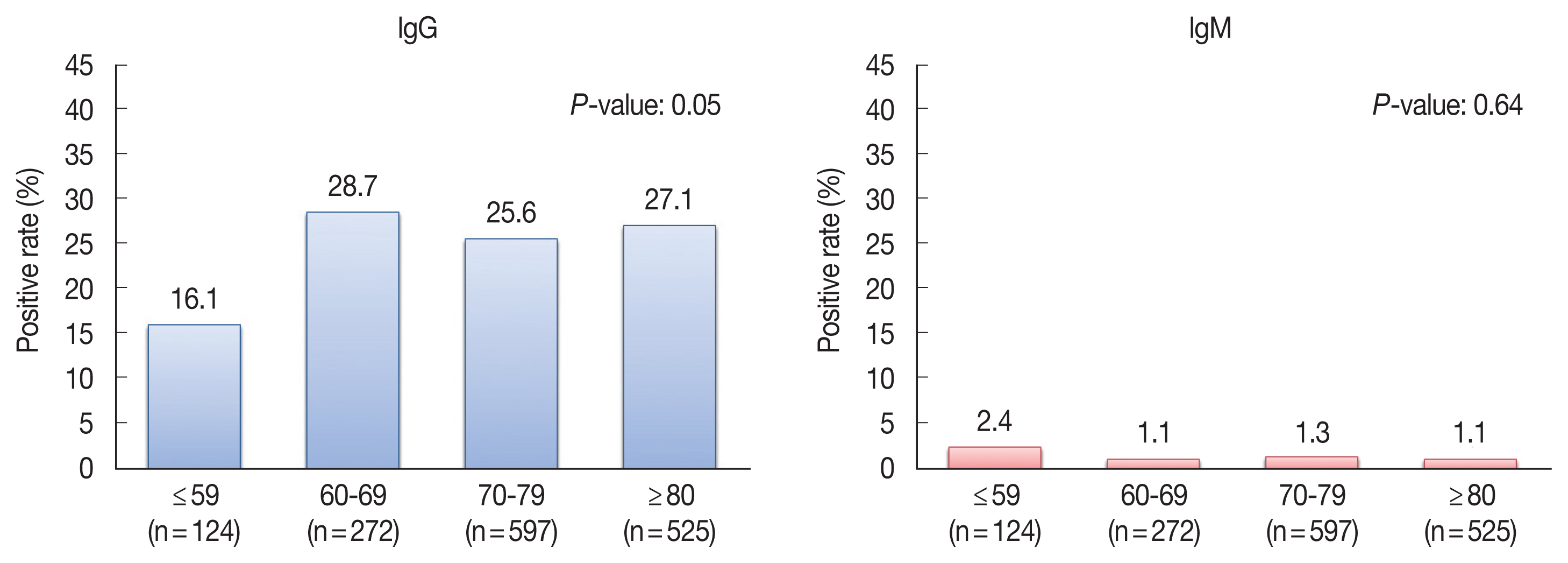
Of the 1,518 overall population, 25.9% of participants were IgG positive while only 1.3% were IgM positive (Table 1). 525 male and 993 female participants were enrolled in this study. 28.4% of men and 24.6% of women were IgG positive (Table 1). 28.1%, 19.5% and 35.7% were IgG positive from Ganghwa, Cheorwon and Goseong respectively. In terms of age groups, 124 were aged less than or equal to 59, 272 were 60 to 69, 597 were 70 to 79 and 525 were older than 80. 16.1%, 28.7%, 25.6%, and 27.1% were IgG positive from age groups less than or equal to 59, 60 to 69, 70 to 79, and 80 plus respectively. The difference among the age groups was not statistically significant; yet, it was very close to 0.05 (Fig. 1). IgM positive rates were from minimum 1.1% to maximum 2.4%. Since IgM positive rate is very low, it was hard to detect statistical difference with current sample size.
Among the 1,518 participants, 552 were from Ganghwa; 661 and 305 were from Cheorwon and Goseong respectively. In Ganghwa, 190 men and 362 women were surveyed, 20 were aged less than or equal to 59, 90 were aged between 60 and 69, 219 were between 70 and 79 and 223 were 80 or older. IgG seroprevalence was 39.5% in men and 22.1% in women. In Cheorwon, 254 men and 407 women were surveyed; among them, 92 were aged 59 or under, 141 were 60 to 69, 251 were 70 to 79 and 177 were 80 or older. IgG seroprevalence in Cherowon was 19.7% in men and 19.4% in women. 81 men and 224 women were surveyed in Goseong, of which 12 were aged 59 or younger, 41 were aged 60 to 69, 127 were aged 70 to 79 and 125 were aged 80 or older. IgG seroprevalence was 29.6% in men and 37.9% in women (Tables 1, 2). Age group less than or equal to 59 years old showed lower incidence rate than other age groups, but no apparent increasing trend was shown as age increases (Table 2). In case of IgM seroprevalence, it appeared from 0% to 3.9%; the numbers of cases were very small (Tables 1, 2). In all 3 study areas, men had higher seroprevalence than women, and more than 50% of the population were elders over the age of 70. Ganghwa has the highest male IgG seroprevalence rate (39.5%), while Goseong has the highest female prevalence (37.9%).
Logistic Regression
Odds can be defined as a chance or likelihood of a certain event or a disease occurs. In a logistic regression model, an odds ratio (OR) represents a level of association between a risk factor and a disease adjusting other variables that were included in the model. If OR equals to 1, it means that there’s no association between the risk factor and the disease. When OR is greater than 1, the risk group has higher odds of the disease occurrence than the non-risk group. Similarly, OR less than 1 can be interpreted that the risk group has lower odds of the disease occurrence than the non-risk group. In Table 3, if we suppose Choerwon as a risk group and Ganghwa as a comparison group (non-risk group), then odds of IgG T. gondii occurrence in Choerwon is 0.63 times lower than that in Ganghwa (OR 0.63, 95% CI 0.48–0.83, P-value 0.001) adjusting age and sex. Using the same interpretation, OR of Goseong versus Ganghwa was 1.47 (95% CI 1.09–1.99, P-value 0.01), and we can conclude that odds of T. gondii event in Goseong is 1.47 times higher than that in Ganghwa. Since P-values of both ORs were less than the statistically significant level of 0.05, seroprevalence of T. gondii in Goseong is statistically higher than that in Ganghwa while T. gondii odds in Cheorwon is statistically lower than that in Ganghwa. Goseong not only has the highest prevalence rate in Table 1 but also has the highest odds in the statistical analysis. Yet, there was no regional difference found with the IgM model. In terms of gender difference in T. gondii infection, men have statistically higher odds of T. gondii by IgG compared to women (OR 1.34, 95% CI 1.05–1.71, P-value 0.02) adjusting region and age; no difference found with IgM. Age group of 60 to 69 had higher odds than the age group of 59 or younger (OR 1.96, 95% CI 1.13–3.42, P-value 0.02) in IgG, but other age group comparisons were not statistically significant. There was no evident age effect on T. gondii prevalence in both IgG and IgM. The IgM test showed no difference in T. gondii seroprevalence for sex, region and age, while IgG method had statistically significant regional and gender effect on T. gondii prevalence.
DISCUSSION
Soh et al. [21] first reported T. gondii seroprevalence of 5.6% among 373 participants in 1960s using toxoplasmin skin test. Since then, various serological methods, indirect fluorescent antibody test (IFAT), indirect latex agglutination test and ELISA, were used. However, these methods were time-consuming and expensive. In this study, Rapid diagnostic test (RDT) using recombinant protein as an antigen was performed which could save time and expenses.
The prevalence of T. gondii in Korea has remained relatively low since the first survey in 1960 until 2010. The 2010 study in Ganghwa and Cheorwon showed a sharp increase in prevalence. Such surge could be related to environmental and socioeconomic changes in Korea. As the Korea economy has grown rapidly, interest in health and environmental issues naturally began to grow. The use of pesticides decreased due to popularity of organic foods, which possibly could lead to increase in exposure to T. gondii [22,23].
Geographical factor also could be contributed to the increase in seroprevalence. Ganghwa, Cheorwon and Goseong are DMZ areas which have the zones that ban civilian access. Due to such characteristic, higher number of wild animals inhabit in these areas compared to other parts of Korea, and the wild animals are the potential reservoir hosts of T. gondii.
According to studies conducted in Ganghwa and Cheorwon from 2010 to 2013, men’s prevalence in both regions was 1.5 times higher than that of women [17,18]. Previous studies have speculated that this phenomenon is due to the fact that men are more active in socioeconomic activities than women in rural areas. Also, some elders in rural, especially in men, believe that consuming animal’s intestines raw is good for their health. In this 2019 study, men had a higher prevalence rate in Ganghwa, but women had a higher prevalence rate in Goseong. Yet, men had statistically significantly higher odds than women adjusting region and age in the logistic regression model.
Another reason for the surge in the T. gondii seroprevalence is the increase in meat consumption. Consuming undercooked meat that consists T. gondii as cyst can cause the infection. The annual meat consumption in Korea increased by more than 20 kilograms from 31.9 kilograms per person in 2000 to 53.9 kilograms per person in 2018. And, meat imports increased 114% from 394,000 tons in 2000 to 845,000 tons in 2017 to meet the demand. T. gondii is widespread around the world, but there is a risk of infection, especially if meat is imported from countries such as Argentina and Brazil, which have high prevalence rates. Furthermore, no quarantine is currently being carried out on T. gondii in imported meat.
Another cause of the spike in prevalence can be attributed to the increase in pet popularity. Pet cats are known to be less likely to be a source of infection for T. gondii; yet, they can become infectious agents if they contact with infected wild animals. Pets in rural areas have more chance to contact with wild animals, which can be thought of as one of reasons for the high seroprevalence rate in rural areas. As cats become popular as pets, the number of abandoned cats also increases which led to the increase in the number of feral cats. The T. gondii infection rate was 38.9% in 72 feral cats in 9 districts of Seoul in 2010. In 2011, 456 wild cats in 17 districts in Seoul were tested and infection rate was about 16%.
The first thing to note in this study is the decrease in prevalence in Cheorwon and Ganghwa. The prevalence rate in Cheorwon was on the rise from 19.3% in 2010 to 26.8% in 2013; however, in 2019, the rate dropped to 19.5% which is similar to the level in 2010. In the case of Ganghwa, the rate increased steeply for 3 consecutive years from 21.7% in 2010 to 33.1% in 2013 but decreased to 28.1% in 2019. The most likely cause of this decrease in prevalence is the change in age groups. The prevalence rate was the highest among those aged 80 or older in 2013; as time goes by, the death of those elders in 2013 could bring a decrease in the overall prevalence rate in 2019. Considering that the prevalence rates in the 60s are not significantly different comparing 2013 and 2019, except for the effect of reducing the prevalence rate caused by deaths in elders, the prevalence rate is on a stabilized trend. For the low prevalence rate in 50s, we should consider that the investigated areas were rural and the number of aged 59 or younger were only 124 combining all 3 areas which is very low number compared to other age groups.
Unlike Ganghwa and Cheorwon, Goseong was first investigated in this study and had a high prevalence rate of 35.7%. Goseong’s prevalence rate is higher than the highest rate of 33.1% in 2013 Ganghwa. In addition, statistical analysis also showed Goseong had higher odds than in other regions. Goseong was expected to show a similar prevalence rate as Cheorwon since both has similar geographical characteristics; however, the prevalence rate in Goseong was much higher than that of Cheorwon. Therefore, further investigation is needed whether other DMZ areas also have such high prevalence rate.
Toxoplasmosis is widespread around the world, but it has been neglected in health policies as the symptoms are often mild or asymptomatic. Since T. gondii prevalence rate before 2010 in Korea was estimated around 10%, which was much lower than the global level of 25 to 30%, it was not considered as an important issue. However, the prevalence rate of Ganghwa, Cheorwon and Goseong in 2019 is 25.9% similar to the global level. Because the investigated areas are rural regions, the estimated prevalence is likely to be higher than that of other regions in Korea, but it is hard to say that this infection problem is only limited to rural areas. Toxoplasmosis can cause serious complications such as encephalitis and ocular toxoplasmosis when reactivated in immunodeficiency. Infection in a pregnant woman can cause a maternal transmission, leaving a neurological after effect. These clinical significances have been ignored, but with the growing elderly population, the need for related research will grow even more. As this study confirmed that the high prevalence of T. gondii continues in some parts of Korea, nationwide prevalence investigations and appropriate health policies are needed.