INTRODUCTION
Feral cats are known to be frequently infected with a host of parasite species. Some cat parasites can infect humans, and can function as the causative agent of zoonosis. In Korea, cats are often reared at home as a pet and/or exploited as a predator of rats. However many of them become feral or stray cats as the result of changes in housing patterns. These cats live freely in urban and rural areas, and tend to discharge helminth eggs, larvae, and protozoan cysts into the general environment (Jang, 1975; Min, 1981; Uga et al., 1989; Oikawa et al., 1991; Saito et al., 1995). Accordingly, feral cats are important as the potent reservoir hosts of a variety of parasites in medical and veterinary point of view.
Many surveys regarding cat helminth have been conducted in Korea (Kang, 1967; Lee 1979; Huh et al., 1993; Yang et al. 1995), Japan (Uchida et al., 1982; Uga et al., 1983; Tanaka et al., 1985; Asato et al., 1986) and other countries (Ash, 1962; Rao and Anantaraman, 1967; Nichols et al., 1981a & b). In Korea, Kang (1967) examined 51 cats obtained in the western part of Kyongsangnam-do. Subsequently, Lee (1979) evaluated the status of 65 cats obtained from Kyongsangbuk-do, Huh et al. (1993) examined 41 cats which had been purchased in a market of Seoul, and Yang et al. (1995) conducted a study with 133 cats from Iri-shi, Chollabuk-do. The numbers of cats examined in these studies were generally small, though, and the cats were collected from fairly restricted localities. The objective of the present study, therefore, was to determine the helminthic infection status of a group of feral cats collected from certain localities within the Republic of Korea.
MATERIALS AND METHODS
We purchased the entire viscera of 438 feral cats from a wholesale house of animals located in Kupo, Puk-gu, Busan, Korea, between February 1996 and January 1997. These cats had been caught primarily in the southern regions of Korea, but were also collected from several other localities. The internal organs of the cats (stomach, intestine, lungs, heart, and liver) were examined thoroughly with the naked eye. The small intestine, in particular, was opened longitudinally with a pair of scissors in 0.85% saline, and washed with the same solution until the supernatant had cleared. The intestinal sedimentary contents were then carefully assessed with the naked eyes, as well as under a stereomicroscope. In order to determine the status of Trichinella larval infections, we minced diaphragm tissue with a mortar and pestle, followed by artificial digestion with pepsin-HCl solution in an incubator at 36℃ for 3 hours. The digested material was then washed with saline until the supernatant had cleared, and examined via stereomicroscopy. The stool samples from the recta of the cats were examined by formalin-ether sedimentation technique.
In order to visualize the morphology of the trematodes in detail, we fixed the recovered worms with 10% neutral buffered formalin under slight pressure of a cover glass. The fixed worms were then stained with Semichon's acetocarmine, and measured using a micrometer (OSM-4, Olympus Co.).
RESULTS
Worm collection
We ultimately detected more than 29 helminth species from the 438 feral cats. The majority of the detected species were identified as trematodes, including Clonorchis sinensis, Paragonimus westermani, Eurytrema pancreatitum, Pharyngostomum cordatum, Metagonimus spp., Heterophyes nocens, Pygidiopsis summa, Heterophyopsis continua, Stictodora fuscata, Stictodora lari, Acanthotrema felis, Stellantchasmus falcatus, Centrocestus armatus, Procerovum varium, Cryptocotyle sp., Echinostoma revolutum, Echinostoma hortense, Echinochasmus japonicus, Stephanoprora sp., Plagiorchis muris, Neodiplostomum sp. and diplostomulum (mesocercaria of Diplostomum sp.). We also detected many worms belonging to the Nematoda and Cestoda, including toxocarids, hookworms, Anisakis simplex larvae, Spirometra erinacei, Taenia taeniaeformis, and one tapeworm remains to be identified. One species of Acanthocephala, Bolbosoma sp., was also detected in the small intestines of 2 cats (Table 1).
Toxocarids were the most frequently detected organism. These were detected in the small intestines of 157 cats (35.8%). T. taeniaeformis (33.8%), S. erinacei (28.5%), H. nocens (24.2%) and P. summa (21.0%) were relatively prevalent in the samples, and the rate of infection with other worms is shown in Table 1.
Among the 438 cats examined in this study, 375 (85.6%) were found to have been infected with 1-8 helminth species. The highest frequency of infection was determined to exist in the group infected with 2 species (24.7%), followed by the group of cats infected with only one species- (23.1%), and then 3 species-(18.0%), 4 species- (9.4%), 5 species- (7.8%), 6 species-(1.6%), 7 species- (0.9%), and the 8 species-infected groups (0.2%) (Table 2).
Stool examination
Eggs from at least 12 helminth species were detected in 438 fecal samples. The eggs of S. erinacei (41.3%) were most frequently detected, and those of the toxocarids (24.4%), T. taeniaeformis (14.6%), P. cordatum (14.4%), heterophyid flukes (13.7%), C. sinensis (11.6%), Capillaria hepatica (2.5%), hookworm (2.1%), echinostomatid flukes (2.1%), P. westermani (0.2%), Hymenolepis. diminuta (0.2%) and diphyllobothriid tapeworm (0.2%) were also detected at varying frequencies (Table 1).
Among the 438 examined fecal specimens, 311 (71.0%) were determined to be positive for 1-5 types of helminth egg. The majority of the cats fell into the group that was positive for one type of eggs (32.4%), followed by the totally egg-negative group (29.0%), the group harboring 2 egg types- (25.30%), the 3 type group (9.6%), the 4 type group,- (3.0%) and the group of cats positive for 5 types of eggs (0.7%) (Table 2).
Worm burdens in the infected cats
Out of 157 cats infected with toxocarids, 129 (82.2%) were infected with 1-5 worms (Table 3). A total of 33 cats (7.5%) was confirmed to be infected with hookworms. Most of them (90.9%) were also determined to harbor worm burdens of 1-5 (Table 4).
A total of 24,104 heterophyid flukes (representing at least 11 species) was collected from 384 cats (87.7%). H. nocens was detected in 106 cats (24.2%), and the worm burden in these cats ranged from one to 389 (38.2 in average). The second frequently encountered species was P. summa (21.9%), and the number of worms per infected cat was 70.4 in average. Metagonimus spp. were associated with the most severe infections in the infected cats. Examples of this species were recovered in burdens from one to 3,984 (148.7 in average) from 78 cats (17.8%). Examples of H. continua were collected from 58 cats (13.2%), and the number of worms in each cat ranged from one to 135 (12 in average). Data regarding infections with other heterophyid flukes is shown in Table 5. The worm burdens of 4 heterophyid flukes, namely, H. nocens, P. summa, Metagonimus spp., and H. continua are shown in Table 6. The majority of the cats were infected with between 1 and 50 worms.
Information regarding infections with echinostomatid flukes is provided in Table 7. E. japonicus was collected from 18 cats (4.1%), and the numbers of worms found ranged from one to 146 (22.8 in average). Information regarding infections with other intestinal flukes, with the exception of the Heterophyidae and Echinostomatidae, is provided in Table 8. P. cordatum was detected in 53 cats (12.1%). As a great number of worms was found to have been embedded deeply in the intestinal wall, it proved quite difficult to precisely enumerate the exact number of P. cordatum worms.
Morphologies of the detected eggs
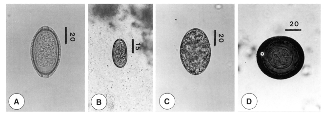
The eggs of C. hepatica were measured to be 57 (51-58) by 31 (19-33) µm in size, and exhibited a thick shell and two mucoid plugs on both poles (Fig. 1). Some of the heterophyid fluke eggs were measured to be 35 (33-37) by 21 (20-22) µm in size, and exhibited miracidia, smooth shells, and faint opercula (Fig. 2). The diphyllobothriid tapeworm eggs were 63 (58-69) by 44 (43-47) µm in size, bilaterally symmetrical, and exhibited weakly developed opercula (Fig. 3). The eggs of H. diminuta were 69 (67-72) by 64 (62-67) µm in size, and possessed oncospheres and thick shells (Fig. 4).
DISCUSSION
Several workers have previously reported on helminth infections in cats in Korea (Kang, 1967; Lee 1979; Eom et al., 1985; Huh et al., 1993; Yang et al. 1995). Kang (1967) detected 5 helminth species, namely, T. cati, C. sinensis, Paragonimus sp., Spirometra sp. and T. taeniaeformis, in 51 cats from the western region of Kyongsangnam-do. Lee (1979) reported detecting 6 trematode species, namely, C. sinensis, H. nocens, M. yokogawai, Centrocestus sp. Echinochasmus perfoliatus, and Echinoparyphium sp., in 65 cats acquired from Kyongsangbuk-do. Eom et al. (1985) described 3 heterophyid flukes, H. continua, H. nocens, and P. summa, which had been collected from 181 domestic cats purchased at the Chungang Market in Seoul. Huh et al. (1993) detected 7 helminth species, namely, T. cati, A. simplex larvae, C. sinensis, P. cordatum, S. erinacei, and T. taeniaeformis, from 41 cats which had been purchased at a market in Seoul. Yang et al. (1995) detected 4 helminth species T. cati, Diphyllobothrium latum, S. erinacei and T. taeniaeformis in 133 cats obtained from Iri-shi, in Chollabuk-do.
A total of 15 helminth species (2 nematodes: T. cati, A. simplex larva; 10 trematodes: C. sinensis, Paragonimus sp., M. yokogawai, H. nocens. H. continua, P. summa, Centrocestus sp., E. perfoliatus, Echinoparyphium sp., P. cordatum; 3 cestodes: S. erinacei, D. latum, T. taeniaeformis) has been designated, thus far, as cat parasites commonly found in Korea. However, according to our results, more than 17 species (1 nematode: Ancylostoma sp.; 15 trematodes: E. pancreatitum, Metagonimus spp., S. fuscata, S. lari, A. felis, S. falcatus, C. armatus, P. varium, Cryptocotyle sp., E. revolutum, E. hortense, E. japonicus, Stephanoprora sp., P. muris and Neodiplostomum sp.; 1 acantocephala: Bolbosoma sp.) should be added to this list. By way of comparison, in Japan, a total of 17 species (6 nematodes: T. cati, Ancylostoma tubaeforme, Strongyloides sp., Dirofilaria immitis, Mammonogamus auris, Oslerus sp.; 6 trematodes: M. yokogawai, P. cordatum, Paragonimus miyazakii, P. westermani, Echinostoma sp., Concinnum okinawanum; 4 cestodes: S. erinacei, T. taeniaeformis, D. caninum, sparganum; 1 acanthocephala: Centrorhynchus sp.) has been isolated from cats (Uga et al., 1983 & 1986; Tanaka et al., 1985; Asato et al., 1986).
Most of the helminth species detected in this study are of great epidemiological importance, as they are zoonotic parasites. In particular, toxocarids, the most frequently encountered species in Korea, are known agents of visceral larva migrans in humans, and S. erinacei is the adult stage of sparganum, which causes human sparganosis. Both of these helminthes can result in very important and possibly serious human diseases. Many trematode species, including 3 major human-infecting trematodes in Korea, i.e., C. sienensis, P. westermani, and M. yokogawai, were detected during this study. Among these, many intestinal trematodes have been reported as the human infecting species in Korea (Chai and Lee, 2002). Our findings suggest that feral cats had a chance to eat the flesh of freshwater fishes, brackish water fishes, and freshwater crabs or crayfish, and they play an important role as reservoir hosts of trematodes.
Helminth infection status in feral cats appears to differ according to the habitats of the cats, i.e., urban or rural, coastal or inland, plains or mountains. Currently, little information is available regarding the precise habitats of the examined cats. However, from the high prevalence of heterophyid flukes, we surmise that many of the cats examined in this study had come from coastal and/or island areas.
There have previously been several helminthological surveys conducted via stool examination (coproscopy) in order to characterize the parasitic infection status of cats in Korea and Japan (Min, 1981; Oikawa et al., 1991; Huh et al., 1993; Saito et al., 1995). Min (1981) detected eggs from 6 helminth species, namely T. cati (7.7%), A. tubaeforme (3.1%), C. sinensis (1.9%), P. westermani (1.4%), M. yokogawai (1.2%) and Spirometra sp. (0.7%) in 416 fecal samples collected from all around the Republic of Korea, and Huh et al. (1993) also detected 6 species, T. cati (41.4%), C. sinensis (12.2%), Metagonimus spp. (9.8%), P. cordatum (7.3%), S. erinacei (41.5%) and T. taeniaeformis (24.4%), from 41 fecal samples obtained from cats that were purchased at a market in Seoul. In this study, we detected more than 12 types of helminth eggs from 438 fecal samples. To our knowledge, eggs of C. hepatica, echinostomes, and H. diminuta had never before been reported in Korean cats. However discharge of C. hepatica eggs via cat feces is originated in the predation of infected rats, and it is epidemiologically important as the one of transmission factors in this nematode infection. Oikawa et al. (1991) reported on the prevalence of intestinal parasites in 1,064 fecal samples from stray cats which had been collected in the western regions of Japan, from 1983 to 1990. Saito et al. (1995) examined 500 fecal samples from cats kept as pets in Fukuyama City, Hiroshima Prefecture, Japan, in 1972 and 1992.
A comparison of helminthic infections examined via two methods (worm collection during autopsy and coproscopic egg detection) was conducted for each parasite species by Nichols et al. (1981); T. cati was detected in 52.2% of autopsies and 36.2% of coproscopies, D. caninum 30.4% and 7.3%, T. taeniaeformis 15.9% and 2.9% and Toxascaris leonina 1.5% and 0%, respectively. Generally, as was reported by Nichols et al. (1981), the detection rate by coproscopy was far lower than that by autopsy. However, the rates of coproscopic detection of S. erinacei, C. sinensis and P. cordatum were far higher than associated with autopsy in this study; S. erinacei was detected in 28.5% of autopsies and 41.3% of coproscopies, C. sinensis was detected at frequencies of 5.5% and 11.6%, and P. cordatum was detected in 12.1% of autopsies, and 14.3% of coproscopies. This trend was also seen with the detection rates of toxocarids, Ancylostoma sp., and T. taeniaeformis via coproscopy and autopsy in this study; toxocarids were detected by autopsy at a rate of 35.8% and by coproscopy at a rate of 24.4%, Ancylostoma sp. were detected at rates of 7.5% and 2.1%, and T. taeniaeformis was detected at rates of 33.8% by autopsy, and 14.6% by coproscopy.
Recent trends of parasitic infections in Korea are typified by remarkable reductions in the frequency of soil-transmitted nematodiasis, the moderate endemicity of foodborne trematodiasis, a gradual increase in zoonosis, an increase in the detection of ostensibly imported parasitosis and oppotunistic infections, and the reemergence of malaria (Lee et al., 1996; MHSA and KAH, 1997). It is worthy of mention that feral cats function as reservoir hosts for zoonosis and foodborne trematodiasis. Nowadays, the importance placed on the incidence of these parasitic infections is steadily increasing. In this study, we confirmed that feral cats in Korea tend to be infected with many different helminth species, which tend to induce zoonosis. Among these species, E. pancreatitum, S. fuscata, S. lari, A. felis, S. falcatus, C. armatus, P. varium, P. muris, E. revolutum, E. japonicus, Cryptocotyle sp., Stephanoprora sp., Neodiplostomum sp. and Bolbosoma sp. are all helminth fauna newly reported in feral cats from the Republic of Korea.