Warning: mkdir(): Permission denied in /home/virtual/lib/view_data.php on line 81

Warning: fopen(upload/ip_log/ip_log_2024-04.txt): failed to open stream: No such file or directory in /home/virtual/lib/view_data.php on line 83

Warning: fwrite() expects parameter 1 to be resource, boolean given in /home/virtual/lib/view_data.php on line 84
An aural myiasis case in a 54-year-old male farmer in Korea
| Home | E-Submission | Sitemap | Contact us |  
top_img
Korean J Parasito Search

CLOSE

Korean J Parasito > Volume 37(1):1999 > Article
Cho, Kim, Cho, Huh, and Ree: An aural myiasis case in a 54-year-old male farmer in Korea

Abstract

A 54-year-old male farmer residing in Chunchon, Korea, complaining of blood tinged discharge and tinnitus in the left ear for two days, was examined in August 16, 1996. Otoscopic examination revealed live maggots from the ear canal. The patient did not complain of any symptoms after removal of maggots. Five maggots recovered were identified as the third stage larvae of Lucilia sericata (Diptera: Calliphoridae). This is the first record of aural myiasis in Korea.

The aural myiasis is not a common infestation in Korea. Although it is not a common manifestation in the field of otorhinolaryngology, the possibility of its occurrence always exists. The clinical spectrum was wide from maggots in the ear to otalgia, otorrhea, perforation of drum, bleeding, itching, roaring sound, tinnitus, furuncle of the external ear, and restlessness. Referral of species identification of maggots by clinicians are necessary for the collection of epidemiological data of aural myiasis cases. We report the larval infestation of Lucilia sericata in the left external auditory canal of a man in Chunchon, Korea. This case is the first written report of aural myiasis in Korea.
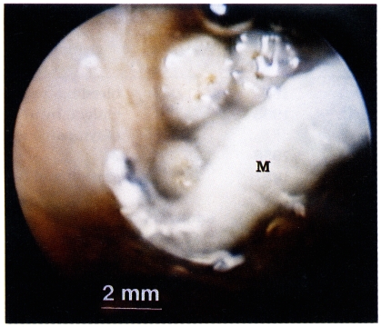
A 54-year-old male farmer visited the outpatient clinic of the Department of Otorhinolaryngology, Hallym University Hospital in August 16, 1996. He complained of blood tinged discharge and tinnitus for two days. Otoscopic examination revealed a number of maggots, blood tinged discharges, and cerumen in the left external auditory canal (Fig. 1). The external auditory canal showed no hyperemic inflammation and tympanic membrane was intact. There were no maggots or lesions in the right external auditory canal. Maggots were removed under the light microscopic field. The external auditory canal was washed with ether to anesthesize worms. There were no maggots or any discharge in the following examinations in one and three weeks. He no longer complained of tinnitus after treatment.
The size of 5 live maggots removed was 10.0-14.1×2.0-2.5 mm (mean 11.5×2.1 mm). The maggots were identified as the third stage larvae of Lucilia (Phaenicia) sericata Meigen (Diptera: Calliphoridae) according to following criteria: mouthparts with no accessory oral sclerite; cephalopharyngeal skeleton without pigmented area below posterior extremity of ventral cornua; posterior spiracle with complete peritreme enclosing a button; inner tubercles of upper margin of anal segment separated by a distance approximately equal to distance between inner and medial tubercles. Speciemen was deposited in the Systematic Entomology Laboratory, Agricultural Research Service, US Department of Agriculture (identificaton lot number 9807357).
Recently there was a case of the third stage larval infestation of Lucilia sp. in the gastrointestinal system of a 91-year-old man admitted due to cerebral infarct in Inchon, Korea. A total of 45 maggots were recovered for two days through nasogastric tube (Chung et al., 1996). Twenty six cases of aural myiasis had been reported in Japan (Chigusa et al., 1994). From literatures, we could find 19 cases of aural myiasis caused by L. sericata including the present case. Blood tinged discharge, tinnitus, discharge, bleeding, pain, and foul smelling discharge were reported symptoms (Table 1). Aural myiasis, like this case, is easily detectable by otoscopic examination.
The treatment of aural myiasis is simple in early manifestation stage, such as removal of maggots and cleansing lesion with 70% ethanol, 10% chloroform, or normal saline (Keller and Keller, 1970; Mendivil and Shamma, 1979; Braverman et al., 1994). If the tympanic membrane is perforated in aural myiasis case, cleansing with normal saline and concomitant suction would be appropriate. When there was a furuncle, the application of antibiotics ointment after cleansing and intake of antibiotics are recommended. In this case, audiogram was not done. If there was a perforation of the tympanic membrane and hearing loss, the examination of auditory function before and after treatment would be necessary. If it is not treated appropriately, the chronic inflammation of the external auditory canal, chronic otitis media or bony destruction from chronic suppuration may be developed (Turnbull and Franklin, 1942).

ACKNOWLEDGEMENTS

We appreciate Mr. Norman E. Woodley, Systematic Entomology Laboratory, Agricultural Research Service, US Department of Agriculture, Building 046, BARC-W, 10300 Baltimore Avenue, Beltsville, MD 20705, USA for his kind identification of maggot.

REFERENCES

1. Baker MC. Green bottle fly worm infestation of the ear. Laryngoscope 1953;63:545-548. PMID: 13070639.
pmid
2. Braverman I, Dano I, Saah D, Gapany B. Aural myiasis caused by flesh fly larva, Sarcophaga haemorrhoidalis. J Otolaryngol 1994;23:204-205. PMID: 8064961.
pmid
3. Chigusa Y, Tanaka K, Yokoi H, et al. Two cases of otomyiasis caused by Sarcophaga peregrina and S. similis (Diptera: Sarcophagidae). Jpn J Sanit Zool 1994;45:153-157.
crossref
4. Chung PR, Jung Y, Kim KS, Cho SK, Jeong S, Ree HI. A case of internal myiasis in Korea. Korean J Parasitol 1996;34:151-154. PMID: 8925248.
pmid
5. Davies DM. Human aural myiasis: a case in Ontario, Canada, and a partial review. J Parasitol 1976;62:124. PMID: 1255358.
crossref pmid
6. El Deeb NE, El Matarawy OM, Ali AA, et al. Human aural myiasis in Taif, Saudi Arabia. J Egypt Soc Parasitol 1988;18:129-132. PMID: 3373040.
pmid
7. Keller AP, Keller AP. Myiasis of the middle ear. Laryngoscope 1970;80:646-650. PMID: 5440720.
crossref pmid
8. Lee HL. Human myiasis in Australia. Med J Aust 1968;1:170-173. PMID: 5689126.
crossref pmid
9. Markova M, Rozkosny R. Myiasis in the ORL area. Cesk Otolaryngol 1984;33:257-259. PMID: 6488380.
pmid
10. Mendivil JA, El Shamma NA. Aural myiasis caused by Cochliomyia hominivorax: case report. Mil Med 1979;144:261-262. PMID: 108625.
pmid
11. Ohkawa T. Biotic foreign bodies in otolaryngology. Pract Otol Kyoto 1979;72:1105-1175.
crossref
12. Turnbull FM, Franklin LB. Aural infestation with screw worms. JAMA 1942;120:117-119.
crossref
13. Yoshi M, Ohkawa C. A case of otomyiasis caused by Phaenicia sericata. J Otolaryngol Jpn 1957;60:994.

Fig. 1
Lucilia sericata maggot (M) in the left external auditory canal of the male farmer seen via otoscope.
kjp-37-51-g001.jpg
Table 1.
Summary of the aural myiasis cases caused by Lucilia sericata
No. of cases Year of detectioin Age/sex Symptoms & signs Country Reference
1 1952 41/M bleeding, pain USA Baker (1953)
2 1955 74/F otalgia, hyeremia, perforation of tympanic membrane Japan Ohkawa (1979)
1958 25/F ?
1 1957 74/F erosion and hyperemia Japan Yoshii and Ohkawa (1957)
1 1962 adult/M ? Australia Lee (1968)
1 1983 47/F discharge Chekoslovakia Markova and Rozkosny (1984)
1 ? 65/F pain, foul smelling discharge USA Keller and Keller (1970)
1 ? 80/M no abnormality Canada Davies (1976)
10 ? 6-17/M ? Saudi Arabia El Deeb et al. (1988)
1 1996 54/M blood tinged discharge and tinnitus Korea Present case
Editorial Office
Department of Molecular Parasitology, Samsung Medical Center, School of Medicine, Sungkyunkwan University,
2066 Seobu-ro, Jangan-gu, Suwon 16419, Gyeonggi-do, Korea.
Tel: +82-31-299-6251   FAX: +82-1-299-6269   E-mail: kjp.editor@gmail.com
About |  Browse Articles |  Current Issue |  For Authors and Reviewers
Copyright © 2024 by The Korean Society for Parasitology and Tropical Medicine.     Developed in M2PI