Warning: mkdir(): Permission denied in /home/virtual/lib/view_data.php on line 81
Warning: fopen(upload/ip_log/ip_log_2024-04.txt): failed to open stream: No such file or directory in /home/virtual/lib/view_data.php on line 83
Warning: fwrite() expects parameter 1 to be resource, boolean given in /home/virtual/lib/view_data.php on line 84 A Clonorchis sinensis-specific antigen that detects active human clonorchiasis
A Clonorchis sinensis-specific antigen that detects active human clonorchiasis
S I Kim
Department of Parasitology, Chosun University College of Medicine, Kwangju 501-759, Korea.
Received November 24, 1997; Accepted January 21, 1998.
Abstract
A Clonorchis sinensis-specific antigen in excretory-secretory product of C. sinensis (CsE) was assessed in human clonorchiasis by immunoblot. Thirty and 7 kDa antigens of CsE2, one of four different batches of CsEs reacted strongly with infection sera from clonorchiasis patients; however, the antigens reacted weakly with 6-month post-treatment sera from praziquantel-cured cases, but were still highly detected by the sera from praziquantel-failed patients, indicating that the 30 and 7 kDa antigens can detect antibodies during an active infection. The 30 kDa antigen showed some cross reactions with sera from patients with Paragonimus westermani and Metagonimus yokogawai, while the 7 kDa antigen did not, suggesting that the 7 kDa antigen has high specificity. The 30 kDa antigen reacted with some past clonorchiasis sera, whereas the 7 kDa antigen did not, supporting that antibodies to the 7 kDa antigen are not present in sera from past clonorchiasis patients. In an endemic area, 92% (23/25) of active clonorchiasis patients and 91% (10/11) of mixed infection patients with C. sinensis and M. yokogawai had IgG antibodies to the 7 kDa antigen, while 40% (6/15) of past clonorchiasis individuals and 43% (3/7) of metagonimiasis patients cross-reacted to the antigen. These data suggest that the 7 kDa antigen in an excretory-secretory antigen may serve as a marker of an active clonorchiasis with reliable specificities in past clonorchiasis, paragonimiasis and metagonimiasis.
Figures
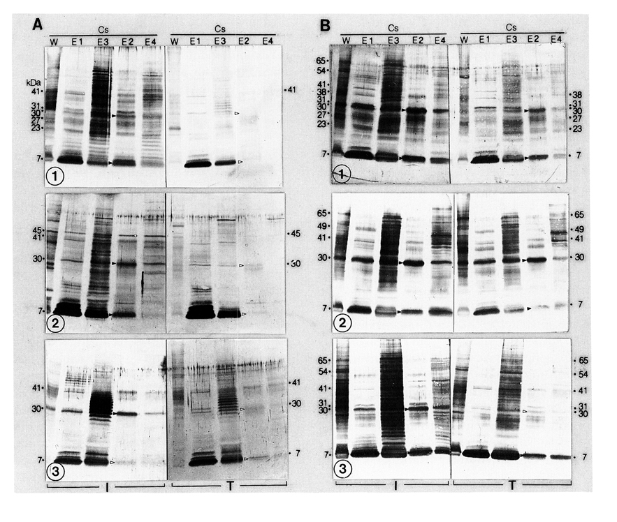
Fig. 1 Immunoblot analyses of CsW and CsE1-4 of C. sinensis probed with paired infection (I) and 6-month post-treatment (T) sera from praziquantel-cured (A) and praziquantel-failed (B) clonorchiasis patients. Amounts of 5 µg protein from each antigen were electrophoresed. Molecular masses in kDa were estimated with standard markers of Bio-Rad. Closed arrow-heads mean 30 and 7 kDa antigen bands that were observed remarkably; open ones mean these bands that were not discernible.
Fig. 2 Immunoblot analyses of CsW and CsE1-4 of C. sinensis reacted with paragonimiasis (A), metagonimiasis (B) and normal control sera (C).
Fig. 3 Immunoblot analyses of CsW and CsE1-4 of C. sinensis reacted with the past clonorchiasis sera (1-5) and conjugate control (6).
Fig. 4 Immunoblot analysis of CsE2 of C. sinensis reacted with paired infection (left strips) and 6-month post-treatment (right strips) sera from praziquantel-cured (A) and praziquantel-failed (B) clonorchiasis patients. Amount of 65 µg protein was electrophoresed in a preparative tract and 40 nitrocellulose strips were prepared.
Fig. 5 Immunoblot analysis of CsE2 of C. sinensis reacted with sera from past clonorchiasis (A), active clonorchiasis (B), metagonimiasis (C), and mixed clonorchiasis and metagonimiasis patients (D).
Tables
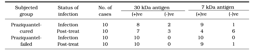
Table 1 Immunoblot analyses of 30 and 7 kDa antigens of CsE2 probed with paired infection and 6-month post-treatment sera from praziquantel-cured and praziquantel-failed clonorchiasis patients
Table 2 Immunoblot analysis of 7 kDa antigen of CsE2 probed with sera from various subjects in an endemic area of clonorchiasis and meta gonimiasis
References
1.
Cho KM, et al. Yonsei Rep Trop Med 1974;7(1):26–39.
2.
Hong ST, Kho WG, Lee M, Lee JS, Lee SH. Immunoblot patterns of clonorchiasis. Korean J Parasitol 1997;35(2):87–93.
3.
Hotez PJ, Zheng F, Long-qi X, Ming-gang C, Shu-hua X, Shu-xian L, Blair D, McManus DP, Davis GM. Emerging and reemerging helminthiases and the public health of China. Emerg Infect Dis 1997;3(3):303–310.
4.
Kim DC, et al. Report NIH, Korea 1969;6:291–308.
5.
Kim J, Chai JY, Kho WG, Cho KH, Lee SH. Immunohistochemical study on the antigenicity of each organ structure of Clonorchis sinensis. Korean J Parasitol 1991;29(1):21–29.
6.
Kim SI. [Immune reactions between excretory-secretory antigens and specific antibodies of Clonorchis sinensis before and after praziquantel treatment in experimentally infected rabbits]. Korean J Parasitol 1994;32(1):35–42.
7.
Lee JK, et al. Yonsei J Med Sci 1981;14(1):133–147.
8.
Min DY, Soh CT. Evaluation Of Specific Ige Antibody In Clonorchis Sinensis Infection. Korean J Parasitol 1983;21(1):27–31.
9.
Min DY, et al. aa. Yonsei J Med Sci 1980;13(2):94–106.
11.
Rim HJ. The current pathobiology and chemotherapy of clonorchiasis. Korean J Parasitol 1986;24 Suppl:1–141.
12.
Rim HJ, et al. New Med J 1973;16(1):69–79.
13.
Sun T, Gibson JB. Antigens of Clonorchis sinensis in experimental and human infections. Am J Trop Med Hyg 1969;18(2):241–252.
14.
Yang TY, et al. aa. Korean J Int Med 1995;49(5):591–597.
15.
Yong TS, Im K, Chung PR. Analysis of Clonorchis sinensis antigens and diagnosis of clonorchiasis using monoclonal antibodies. Korean J Parasitol 1991;29(3):293–310.