Warning: mkdir(): Permission denied in /home/virtual/lib/view_data.php on line 81
Warning: fopen(upload/ip_log/ip_log_2024-04.txt): failed to open stream: No such file or directory in /home/virtual/lib/view_data.php on line 83
Warning: fwrite() expects parameter 1 to be resource, boolean given in /home/virtual/lib/view_data.php on line 84 Expression patterns of proliferating cell nuclear antigen in the small intestine of mice infected with Metagonimus yokogawai and Metagonimus Miyata type
Expression patterns of proliferating cell nuclear antigen in the small intestine of mice infected with Metagonimus yokogawai and Metagonimus Miyata type
J R Yu,*1N Myong,2 and J Y Chai3
1Department of Parasitology, College of Medicine, Kon-Kuk University, Chungju 380-701, Korea.
Received August 25, 1997; Accepted October 06, 1997.
Abstract
Genus Metagonimus has been a subject of taxonomic debates for several years. In morphological aspects, M. yokogawai has been thought to have three subtypes, namely Yokogawa type (M. yokogawai in strict sense), Miyata type and Koga type, But differences in the intestinal pathology induced by these subtypes have not been studied yet. In this study we compared the pathological reactions induced by M. yokogawai and Metagonimus Miyata type using proliferating cell nuclear antigen (PCNA) index. Metacercariae (Mc) of M. yokogawai were collected by artificial digestion of Plecoglossus altivelis and Mc of Metagonimus Miyata type were collected from Zacco platypus. Three hundreds Mc of each species were infected orally to ICR mice. The mice were sacrificed at 3, 6, 10, 16 and 23 days after infection and the small intestines were resected into three portions (proximal, middle, and distal). Immunohistochemical staining for PCNA was done using PC-10 (DAKO-PCNA, CA, USA). The PCNA indices in M. yokogawai infected group on the 6th and 23rd day after infection were lower than in the control and Miyata type infected groups (p < 0.05) from all of the three intestinal regions. On the other hand, the control group and Metagonimus Miyata type infected group did not make any differences in PCNA indices. The villus/crypt (V/C) ratio was also decreased significantly in M. yokogawai infected mice but not in Metagonimus Miyata type infected ones. It is suggested that M. yokogawai induce villous atrophy through a decrease in the cell proliferation at the crypt. The results of this study suggested that M. yokogawai induce more serious intestinal pathology than Metagonimus Miyata type.
Figures
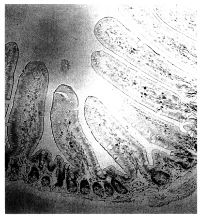
Fig. 1 Preparation of the small intestine of infected mice. P, Proximal small intestine; M, middle small intestine; D, distal small intestine.
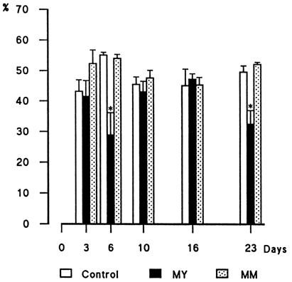
Fig. 3 Comparison of PCNA indices among experimental groups in the proximal jejunum of mice. MY, M. yokogawai infected group; MM, Metagonimus Miyata type infected group. *p<0.05, student's t-test.
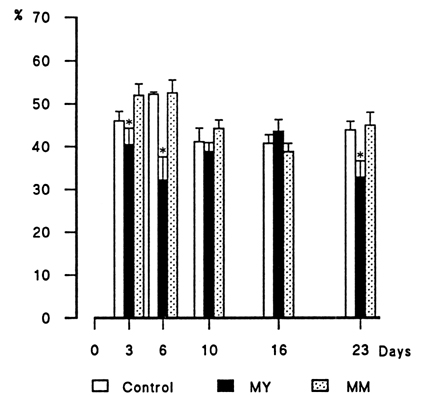
Fig. 4 Comparison of PCNA indices among experimental groups in the middle intestine of mice. MY, M. yokogawai infected group; MM, Metagonimus Miyata type infected group. *p<0.05, student's t-test.
Fig. 5 Comparison of PCNA indices among expermental groups in the distal small intestine of mice. MY, M. yokogawai infected group; MM, Metagonimus Miyata type infected group. *p<0.05, student's t-test.
Tables
Table 1 The number of mice used for this experiment
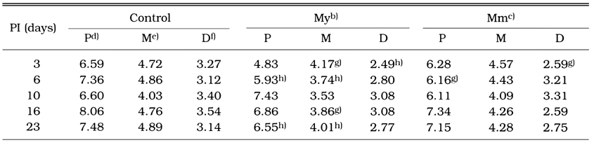
Table 2 Comparative valuesa) of V/C ratio among experimental groups
References
1.
Chai JY, Huh S, Yu JR, Kook J, Jung KC, Park EC, Sohn WM, Hong ST, Lee SH. An epidemiological study of metagonimiasis along the upper reaches of the Namhan River. Korean J Parasitol 1993;31(2):99–108.
2.
Chai JY, Lee SH. Intestinal trematodes of humans in Korea: Metagonimus, heterophyids and echinostomes. Korean J Parasitol 1990;28 Suppl:103–122.
3.
Chai JY, et al. Seoul J Med 1989;30:139–142.
4.
Chai JY, Yun TY, Kim J, Huh S, Choi MH, Lee SH. [Chronological observation on intestinal histopathology and intraepithelial lymphocytes in the intestine of rats infected with Metagonimus yokogawai]. Korean J Parasitol 1994;32(4):215–221.
5.
Saito S. Reports on Meetings for Parasite Taxonomy and Morphyolgy, Japan. 1984. pp. 1-4.
6.
van Dierendonck JH, Wijsman JH, Keijzer R, van de Velde CJ, Cornelisse CJ. Cell-cycle-related staining patterns of anti-proliferating cell nuclear antigen monoclonal antibodies. Comparison with BrdUrd labeling and Ki-67 staining. Am J Pathol 1991;138(5):1165–1172.
7.
Yu JR, Chung JS, Chai JY. Different RAPD patterns between Metagonimus yokogawai and Metagonimus Miyata type. Korean J Parasitol 1997;35(4):295–298.
8.
Yu JR, Chung JS, Huh S, Lee SH, Chai JY. PCR-RFLP pattern of three kinds of Metagonimus in Korea. Korean J Parasitol 1997;35(4):271–276.
9.
Yu JR, Hong ST, Chai JY, Lee SH. The effect of reinfection with Neodiplostomum seoulensis on the histopathology and activities of brush border membrane bound enzymes in the rat small intestine. Korean J Parasitol 1995;33(1):37–43.
10.
Yu JR, Kwon SO, Lee SH. Clonorchiasis and metagonimiasis in the inhabitants along Talchongang (River), Chungwon-gun. Korean J Parasitol 1994;32(4):267–269.