INTRODUCTION
Genus Cochlosoma is a flagellated protozoan parasite associated with enteritis and stunting in turkeys and several species of birds [1-5]. Cochlosoma anatis is the only species described, causing infection in birds. Several case reports and studies suggest that C. anatis is a significant pathogen in both turkeys and ducks [1-3]. Involvement of the protozoan in outbreak of diarrhea in ducks and turkeys has been reported by Cooper et al. [2] and Bolinger and Barker [3]. In many cases, C. anatis was the only pathogen identified; however, bacteria, viruses, or other protozoa may interact synergistically with Cochlosoma infection [1]. In turkey poults, which were inoculated experimentally with C. anatis isolated from natural cases, reduced weight and delayed feathering were observed but diarrhea and other clinical abnormality were absent [6]. The noncyst forming trophozoites of Cochlosoma could transmit the infection to susceptible birds. Since exposure to water or freezing could destroy the trophozoites in a short span of time, contamination of feed or litter with the infected fecal materials would be the major sources of infection for other susceptible birds [6]. In the present study, Cochlosoma infection is reported for the first time in a stunting diarrheic turkey poult in Iran.
CASE DESCRIPTION
A 7-8 week-old native turkey poult (Meleagris galopavo) out of 60 birds showing clinical signs of unthriftness and chronic diarrhea obtained from rural areas in Mazandaran province (one of the northern provinces of Iran) was examined for the causative agent of the disease. The native turkeys were kept as small flocks generally consisting of chickens, turkeys, and waterfowls, and they were kept under free ran condition. The clinical manifestations of the affected bird were recorded and the bird was sacrificed. The tissue samples taken from the duodenum, jejunum, ileum, cecum , colon, cloaca, and bursa of Fabricius were fixed in 10% buffered formalin (pH = 7.2) The formalin-fixed tissues were processed in a tissue processor, paraffin blocks were made, and 5-6 µm thick sections were prepared and stained with Harris hematoxylin and eosine (H-E) method. Several formalin-fixed segments of the duodenum, jejunum, ileum, cecum, colon, cloaca, and bursa of Fabricius were post-fixed in 2.5% glutaraldehyde and processed through standard method for transmission electron microscopy (TEM). The ultrathin sections stained with uranyl acetate and lead citrate were examined under a Philips 208 S transmission electron microscope.
CLINICAL FINDINGS AND PATHOLOGY
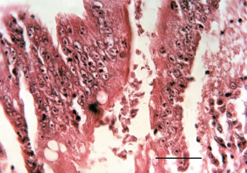
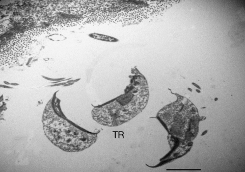
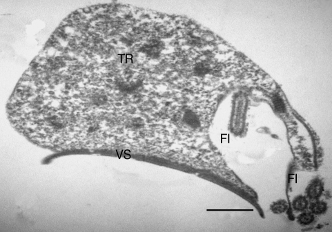
Growth retardation, depression, and chronic watery diarrhea were recorded for the affected turkey poult. In histological examinations, moderate enteritis characterized by blunting and fusion of villi, villous edema, an increase in lamina propria inflammatory cells, and increased number of mitotic figures in the crypt epithelial cells were noticed. Large numbers of flagellated conical protozoan were present within the lumen and intervillous spaces of the jejunum, ileum, and cecum (Fig. 1). Some of which seem to be firmly attached to the gut epithelial lining. In TEM studies on parasagittal sections, the trophozoites of the organism was a uninuclear, conical, or pyriform-shaped flagellated cell with a prominent ventral sucker-like disc (Figs. 2-4). There were sharp points at the posterior and anterior ends of the ventral sucker and 6 flagella emerging from an opening space of ventrodorsal surface of the parasite (Figs. 3, 4). In cross sections, the sucker seemed to be concave and had an electron dense free sharp edge. The morphological characteristics of the organism are in agreement to those described for Cochlosoma anatis.
DISCUSSION
In the present study, a Cochlosoma infection was diagnosed in a native turkey poult. The infection was accompanied by growth retardation and persistent diarrhea. Although the pathogenicity of this protozoan is controversial, several reports from naturally occurring infections and experimental data indicate that the organism could be infective and pathogenic for turkeys and several species of waterfowls [1-5]. Natural infections with C. anatis were reported in turkeys [2] and ducklings [3]. It has also been shown that Cochlosoma could be experimentally infected to turkeys [2,6], and inoculation of turkeys with trophozoites produced severe infections. The localization of the parasite in the infected poult was the jejunum, ileum, and cecum which agrees to those reported in naturally or experimentally infected birds [1-3]. The presence of the parasite in these anatomic sites may be detrimental to the intestinal mucosal function. It seems that mechanical irritation or toxic actions of the parasite upon the gut epithelium or combination of these detrimental effects may attribute to the pathogenicity of Cochlosoma parasite [3]. As proposed, the sharp edges of the ventral sucker may irritate the mucosa at the site of the parasite attachment or during attachment to the gut epithelium. According to Bolinger and Barker [3] and Bermudez [1], only a single species of Cohlosoma, namely C. anatis has been identified to be causing infections in turkeys and several species of water fowls. The characteristic features of C. anatis described [1-5], including pyriform or conical shape, the ventral sucker with sharp edge and the presence of 6 flagella, are compatible with the parasite found in the present case. However, molecular confirmation is required [7].
Several TEM pictures of the parasite made us propose that the ventral sucker may be composed of a relatively rigid structure that partially maintains and supports the morphology of the trophozoite. The ventral sucker seems to be a plate-like structure with an electron-dense margin and a concave center which is composed of a modified cytoplasmic membrane. The cytoplasmic non-electron dense portion of the ventral sucker may have important physiological functions, for example, as a feeder organ of the parasite, and also may serve as an organ that irritates the mucosal epithelial cells. Perhaps, the ventral sucker enables the parasite to obtain its essential nutrients from the oozed or secreted materials coming from the site of attachment. Therefore, it is supposed that the ventral sucker may have specialized functions required for the protozoan survival and pathogenicity. Since the major sources of the organism are contaminated feed and litter, keeping birds in the free ran condition could facilitate transmission of the infection between susceptible birds.
It is concluded that Cochlosoma infection could be one of the protozoan parasite infections in native turkeys of Iran, and that the infection is associated with runting syndrome and diarrhea.