Warning: mkdir(): Permission denied in /home/virtual/lib/view_data.php on line 81
Warning: fopen(upload/ip_log/ip_log_2024-04.txt): failed to open stream: No such file or directory in /home/virtual/lib/view_data.php on line 83
Warning: fwrite() expects parameter 1 to be resource, boolean given in /home/virtual/lib/view_data.php on line 84 Differentiation of Entamoeba histolytica and Entamoeba dispar in cyst-passers by immunoblot
Differentiation of Entamoeba histolytica and Entamoeba dispar in cyst-passers by immunoblot
Mejeong Lee and Sung-Tae Hong*
Department of Parasitology and Institute of Endemic Diseases, Seoul National University College of Medicine, Seoul, 110-799, Korea.
Received November 15, 1996; Accepted November 27, 1996.
Abstract
Differentiation of invasive strains of Entamoeba histolytica according to their pathogenicity has been a topic of long debate, but now the pathogenic species only is regarded as E. histolytica while the non-pathogenic species is E. dispar. The present study applied immunoblot to differentiate infections of the two species among microscopically-detected cyst-passers in Korea. The crude extract of E. histolytica separated in 5-20% gradient gels, revealed many fractions of 94, 81, 71, 50, 44, 38.5, 37.5, 29, 19, and 18 kDa when the cysteine proteinase inhibitor, E64, was supplemented. The serum IgG antibody of proven E. histolytica cases reacted with the antigenic fractions of 117, 110, 99, 68, 66, 60, 54, 52, 46, and 45 kDa. Sera of PCR confirmed 3 cases of E. dispar reacted only to the 117 kDa fraction of the E. histolytica crude extract which was regarded as non-specific. To the antigen of monoxenic E. dispar, sera of E. dispar and E. histolytica cases showed the same immunoblot reactions. The serum IgA antibody reacted with several antigenic fractions of both E. histolytica and E. dispar, but IgM and IgE antibodies showed no reaction to either antigen. Sera of 24 symptomless amebic cyst-passers were screened with the E. histolytica antigen; two were found to be infected by E. histolytica and 22 were by E. dispar. The present findings suggest that in Korea most asymptomatic cyst passers of E. histolytica are carriers of E. dispar. Immunoblot using E. histolytica antigen is a good technique for the differentiation of E. histolytica and E. dispar infections.
Figures
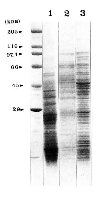
Fig. 1 The effects of proteinase inhibitors on crude extracts of E. histolytica YS-27 trophozoites by SDS-PAGE in a 5-20% gradient gel. Lane 1. serine proteinase inhibitor (PMSF); lane 2, aspartic proteinase inhibitor (pepstatin A); lane 3, cysteine proteinase inhibitor (E64); lane 4, cysteine proteinase inhibitor (iodoacetic acid); lane 5, without proteinase inhibitors.
Fig. 2 Comparison of crude extracts of E. histolytica and E. dispar separated in 5-20% gradient SDS-PAGE. Lane 1, E. histolytica (YS-29); lane 2, E. dispar (S16); lane 3, Escherichia coli.
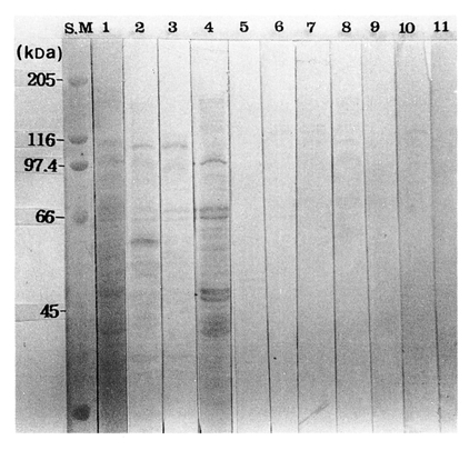
Fig. 3 Immunoblot patterns of serum IgG antibodies to the E64 supplemented crude extracts of E. histolytica. Lane 1, amido black 10B-stained protein fractions; lanes 2-4, sera of E. histolytica patients; lanes 5-8, sera of E. dispar infected humans; lane 9, serum of E. coli infection; lane 10, serum of B. hominis infection; lane 11, serum of normal control.
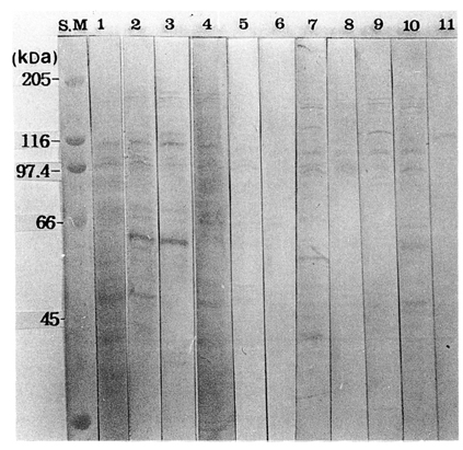
Fig. 4 Immunoblot patterns of serum IgA antibodies to the E64 supplemented crude extracts of E. histolytica. Lane 1, amido black 10B-stained protein fractions; lanes 2-4, sera of E. histolytica patients; lanes 5-8, sera of E. dispar infected humans; lane 9, serum of E. coli infection; lane 10, serum of B. hominis infection; lane 11, serum of a normal control.
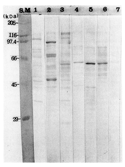
Fig. 5 Immunoblot patterns of serum IgG antibodies to the E64 supplemented crude extracts of E. dispar. Lanes 1-3, sera of E. histolytica infection; lanes 4-6, sera of E. dispar infection; lane 7, serum of a control.
Fig. 6 Immunoblot patterns of serum IgA antibodies to the E64 supplemented crude extracts of E. dispar. Lanes 1-3, sera of E. histolytica infection; lanes 4-6, sera of E. dispar infection; lane 7, control serum.
Fig. 7 Screening of 24 symptomless cyst-passers with antigen of E. histolytica. The sera probed to the strips 11 and 12 were regarded as infection of E. histolytica, and all of ohers were of E. dispar.
References
1.
Abioye AA, Lewis EA, McFarlane H. Clinical evaluation of serum immunoglobulins in amoebiasis. Immunology 1972;23(6):937–946.
2.
Anaya-Velazquez F, Padilla-Vaca F. Effect of intestinal bacteria on the virulence of Entamoeba histolytica. Arch Med Res 1992;23(2):183–185.
3.
Kettis AA, Thorstensson R, Utter G. Antigenicity of Entamoeba histolytica strain nih 200: a survey of clinically relevant antigenic components. Am J Trop Med Hyg 1983;32(3):512–522.
4.
Chang SM, Lin CM, Dusanic DG, Cross JH. Antigenic analyses of two axenized strains of Entamoeba histolytica by two-dimensional immunoelectrophoresis. Am J Trop Med Hyg 1979;28(5):845–853.
5.
Chang JK, Im K, Soh CT. Axenization of Entamoeba histolytica, a Korean strain YS-27. Korean J Parasitol 1995;33(4):387–390.
6.
Choe SC, Lee M, Lee SK, Im K, Tannich E, Lee SH, Hong ST. Differentiation of Korean isolates of Entamoeba histolytica from Entamoeba dispar. Korean J Parasitol 1996;34(1):15–20.
7.
Diamond LS, Clark CG. A redescription of Entamoeba histolytica Schaudinn, 1903 (Emended Walker, 1911) separating it from Entamoeba dispar Brumpt, 1925. J Eukaryot Microbiol 1993;40(3):340–344.
8.
Edman U, Meraz MA, Rausser S, Agabian N, Meza I. Characterization of an immuno-dominant variable surface antigen from pathogenic and nonpathogenic Entamoeba histolytica. J Exp Med 1990;172(3):879–888.
10.
Krupp IM, Powell SJ. Antibody response to invasive amebiasis in Durban, South Africa. Am J Trop Med Hyg 1971;20(3):414–420.
11.
Leammli UK. Nature 1970;227:681–685.
12.
Montfort I, Perez-Tamayo R, Perez-Montfort R, Gonzalez Canto A, Olivos A. Purification and immunologic characterization of a 30-kDa cysteine proteinase of Entamoeba histolytica. Parasitol Res 1994;80(7):607–613.
13.
Ong SJ, et al. Trans Roy Soc Trop Med Hyg 1996;90:248–249.
14.
Sengupta K, Das P, Johnson TM, Chaudhuri PP, Das D, Nair GB. Production and characterization of monoclonal antibodies against a highly immunogenic fraction of Entamoeba histolytica (NIH:200) and their application in the detection of current amoebic infection. J Eukaryot Microbiol 1993;40(6):722–726.
Tannich E, Burchard GD. Differentiation of pathogenic from nonpathogenic Entamoeba histolytica by restriction fragment analysis of a single gene amplified in vitro. J Clin Microbiol 1991;29(2):250–255.
17.
Tsang VC, Peralta JM, Simons AR. Enzyme-linked immunoelectrotransfer blot techniques (EITB) for studying the specificities of antigens and antibodies separated by gel electrophoresis. Methods Enzymol 1983;92:377–391.
18.
Ximenez C, Leyva O, Moran P, Ramos F, Melendro EI, Ramiro M, Martinez MC, Munoz O, Kretschmer R, Arellano J. Entamoeba histolytica: antibody response to recent and past invasive events. Ann Trop Med Parasitol 1993;87(1):31–39.