Warning: mkdir(): Permission denied in /home/virtual/lib/view_data.php on line 81
Warning: fopen(upload/ip_log/ip_log_2024-04.txt): failed to open stream: No such file or directory in /home/virtual/lib/view_data.php on line 83
Warning: fwrite() expects parameter 1 to be resource, boolean given in /home/virtual/lib/view_data.php on line 84 Lymphadenitis in experimental murine toxoplasmosis induced by intramuscular injection of tachyzoites
Lymphadenitis in experimental murine toxoplasmosis induced by intramuscular injection of tachyzoites
Won-Young Choi,*Ho-Woo Nam,Eun-Jung Baek and Seung-Yull Cho
Department of Parasitology, Catholic University Medical College, Seoul 137-701, Korea.
Received May 06, 1995; Accepted May 19, 1995.
Abstract
When tachyzoites (RH strain) of Toxoplasma gondii are injected intramuscularly, experimental mice survive up to 7 days, 1-2 days longer than those infected intraperitoneally. We observed sequential histopathological changes in inguinal lymph nodes after intramuscular injection of tachyzoites to thighs of specific pathogen free (SPF) mice. Initial findings on 1 or 3 days after the injection were reactive germinal centers, distended sinuses and epithelioid cell clusters in cortical and paracortical regions. Later on 5 days after the injection, however, effacement of nodal structure with depletion of cells and focal necrosis were observed. Necrotizing lymphadenitis in the experimental murine toxoplasmosis suggests the causal relation between T. gondii infection and the human disease.
Figures
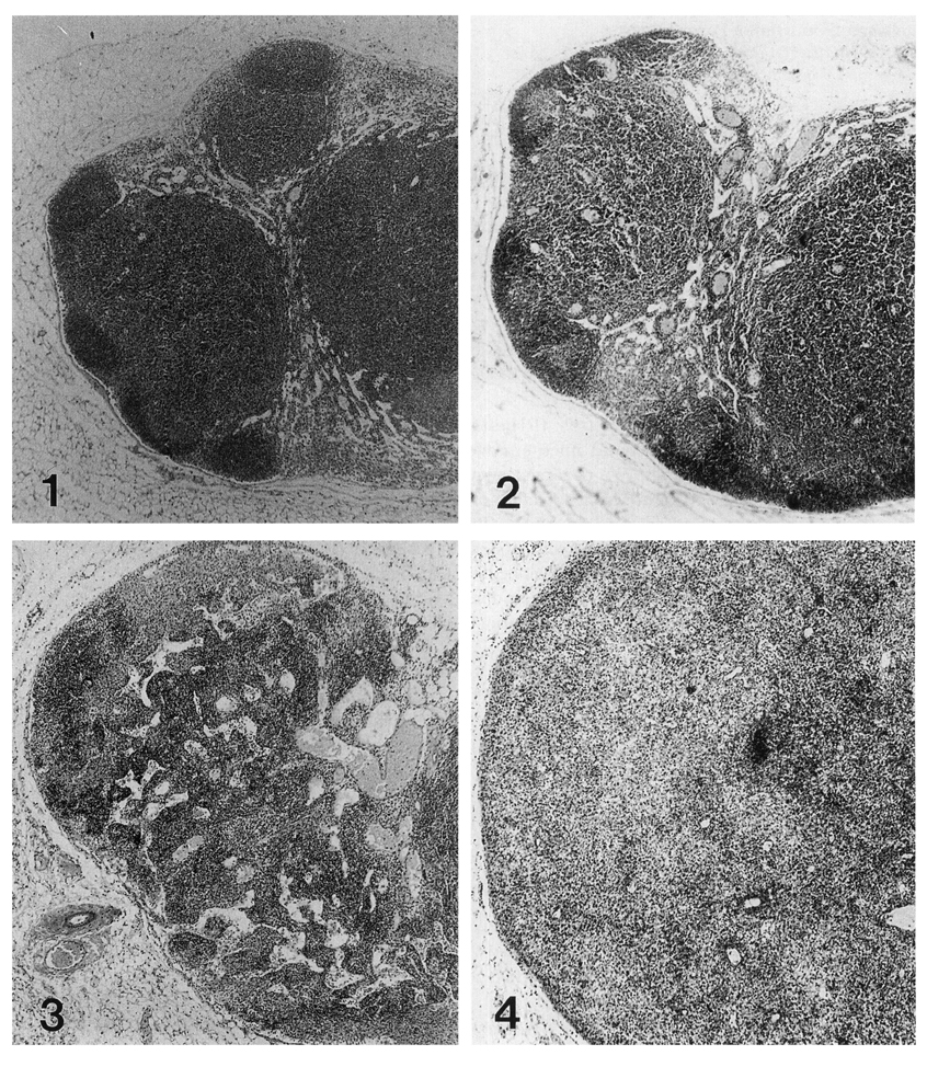
Figs. 1-4 Ipsilateral murine inguinal lymph nodes after the intramuscular injection of tachyzoites to thigh. Hematoxylin-eosin stained, × 40. 1. Control lymph node of non-injected specific pathogen free mouse. 2. Lymph node of one day after the injection. Secondary follicles are well developed in cortical region. 3. Lymph node of 3 days after the injection. Follicular structures disappeared, and lymph sinuses are tortuously expanded and filled with many monocyte-like cells. 4. Lymph node of 5 days after the infection. Lymph nodes are enlarged about 1.5-2 times of non-injected controls. Nodal architecture was diffusely damaged with focal necrosis.
Tables
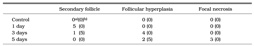
Table 1 The number of ipsilateral and contralateral inguinal lymph nodes of mice which demonstrate specific histopathological findings of toxoplasmic lymphadenitis. Each group consists of 5 mice
References
1.
Cho KJ, Kim CW, Park SH, Lee SK. Necrotizing lymphadenitis--a clinico-pathologic study of 36 cases with immunohistochemical analysis. J Korean Med Sci 1991;6(1):55–61.
2.
Dorfman RF, Remington JS. Value of lymph-node biopsy in the diagnosis of acute acquired toxoplasmosis. N Engl J Med 1973;289(17):878–881.
4.
Jones TC, Kean BH, Kimball AC. Toxoplasmic Lymphadenitis. JAMA 1965;192:1–5.
5.
Kikuchi M. Acta Haematol Jpn 1972;35:379–380.
6.
Kikuchi M, Yoshizumi T, Nakamura H. Necrotizing lymphadenitis: possible acute toxoplasmic infection. Virchows Arch A Pathol Anat Histol 1977;376(3):247–253.
7.
Kim SE, et al. Korean J Infect Dis 1993;25:63–69.
8.
Piringer-Kuchinka A, et al. Virchows Arch A Path Anat Histol 1958;331:552–555.
9.
Saxen E, et al. Acta Pathol Microbiol Scand 1958;44:319–328.