Warning: mkdir(): Permission denied in /home/virtual/lib/view_data.php on line 81
Warning: fopen(upload/ip_log/ip_log_2024-04.txt): failed to open stream: No such file or directory in /home/virtual/lib/view_data.php on line 83
Warning: fwrite() expects parameter 1 to be resource, boolean given in /home/virtual/lib/view_data.php on line 84 Mucosal mast cell responses to experimental Metagonimus yokogawai infection in rats
Mucosal mast cell responses to experimental Metagonimus yokogawai infection in rats
J Y Chai,*1T H Kim,1,**W G Kho,1,**S W Chung,2S T Hong,1 and S H Lee1
Department of Parasitology and Institute of Endemic Diseases, Seoul National University College of Medicine, Seoul 110-799, Korea.
***
Received April 03, 1993; Accepted April 26, 1993.
Abstract
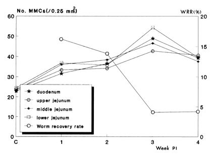
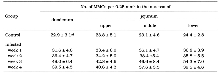
Intestinal mucosal mast cell (MMC) responses were studied in rats experimentally infected with Metagonimus yokogawai (Digenea: Heterophyidae). Twenty Sprague-Dawley rats were fed each 2,500 metacercariae isolated from the sweetfish and sacrificed on the week 1, 2, 3 and 4 post-infection (PI). Recovery of worms was performed from the small intestine of each rat. To visualize the MMCs, duodenal and jejunal (upper, middle and lower) tissue sections were made and stained with alcian blue/safranine-0. The average worm recovery rates were 16.2% and 13.8% on the week 1 and week 2, respectively, but they decreased rapidly to 4.1% and 4.2% on the week 3 and week 4 PI, respectively, which indicate spontaneous worm expulsion after the week 2. The MMC number in the infected rats was, compared with uninfected controls, significantly increased in the whole small intestine, through the whole period of observation. The peak level of mastocytosis was observed on the week 3 PI. It is strongly suggested that MMCs might be involved in the expulsion process of flukes from the rat intestine.
Figures
Figs. 1-4 Sections of the duodenum of rats infected with M. yokogawai in comparison with control group. Blue spots (arrows) in the lamina propria and epithelial layer are the mucosal mast cells (MMCs). 1. uninfected control group, showing only a small number of MMCs. 2. infected rat, week 1 PI, a significant increase in the number of MMCs is recognizable. 3. infected rat, week 3 PI, showing markedly increased number (peak level) of MMCs. 4. infected rat, week 4 PI, with decreased number of MMCs compared with week 3 PI. Alcian blue (pH 0.3)/safranine-0 stainde, × 100.
Fig. 5 Correlations between the worm recovery rate (WRR) and number of MMCs (No. MMCs) in different locations of the small intestine of the rats experimentally infected with M. yokogawai.
Tables
Table 1 Mucosal mast cell (MMC) numbers in the small intestine of rats experimentally infected with M. yokogawai
References
1.
Andreassen J, Hindsbo O, Ruitenberg EJ. Hymenolepis diminuta infections in congenitally athymic (nude) mice: worm kinetics and intestinal histopathology. Immunology 1978;34(1):105–113.
2.
Castro GA. Immunophysiology of enteric parasitism. Parasitol Today 1989;5(1):11–19.
3.
Chai JY. Seoul J Med 1979;20:104–117.
4.
Chai JY, Lee SH. Intestinal trematodes of humans in Korea: Metagonimus, heterophyids and echinostomes. Korean J Parasitol 1990;28 Suppl:103–122.
5.
Guy-Grand D, Dy M, Luffau G, Vassalli P. Gut mucosal mast cells. Origin, traffic, and differentiation. J Exp Med 1984;160(1):12–28.
6.
Hong ST. Studies On Intestinal Trematodes In Korea: VII. Growth, Development And Recovery Of Fibricola Seoulensis From Experimentally Infected Rats And Mice. Korean J Parasitol 1982;20(2):112–121.
7.
Kang SY, Cho SY, Chai JY, Lee JB, Jang DH. A Study On Intestinal Lesions Of Experimentally Reinfected Dogs With Metagonimus Yokogawai. Korean J Parasitol 1983;21(1):58–73.
8.
Kho WG, et al. Seoul J Med 1990;31:191–199.
9.
Lee TD, Swieter M, Befus AD. Mast cell responses to helminth infection. Parasitol Today 1986;2(7):186–191.
10.
Miller HR, Jarrett WF. Immune reactions in mucous membranes. I. Intestinal mast cell response during helminth expulsion in the rat. Immunology 1971;20(3):277–288.
11.
Mimori T, Nawa Y, Korenaga M, Tada I. Strongyloides ratti: mast cell and goblet cell responses in the small intestine of infected rats. Exp Parasitol 1982;54(3):366–370.
13.
Seo BS. Seoul J Med 1990;31:61–96.
14.
Seo BS, et al. Seoul J Med 1971;12:234–241.
15.
Strobel S, Miller HR, Ferguson A. Human intestinal mucosal mast cells: evaluation of fixation and staining techniques. J Clin Pathol 1981;34(8):851–858.
16.
Woodbury RG, Miller HR, Huntley JF, Newlands GF, Palliser AC, Wakelin D. Mucosal mast cells are functionally active during spontaneous expulsion of intestinal nematode infections in rat. Nature 1984;312(5993):450–452.