Warning: mkdir(): Permission denied in /home/virtual/lib/view_data.php on line 81
Warning: fopen(upload/ip_log/ip_log_2024-04.txt): failed to open stream: No such file or directory in /home/virtual/lib/view_data.php on line 83
Warning: fwrite() expects parameter 1 to be resource, boolean given in /home/virtual/lib/view_data.php on line 84 The fate of spargana inoculated into the cat brain and sequential changes of anti-sparganum IgG antibody levels in the cerebrospinal fluid
The fate of spargana inoculated into the cat brain and sequential changes of anti-sparganum IgG antibody levels in the cerebrospinal fluid
K C Wang,*S Huh,**S T Hong,J Y Chai,K S Choi,* and S H Lee
Department of Parasitology, College of Medicine, Seoul National University, Seoul 110-460, Korea.
*Department of Neurosurgery, College of Medicine, Seoul National University, Seoul 110-460, Korea.
***Present address: Department of Parasitology, faculty of Medicine, Hallym University, Chunchon 200-702, Korea.
Abstract
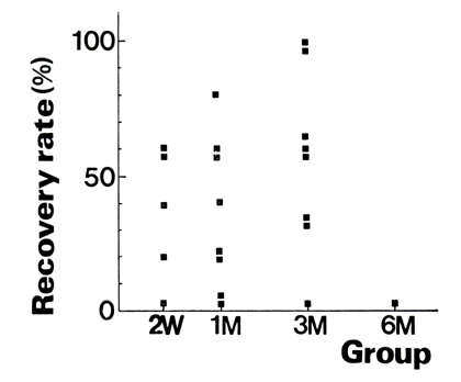
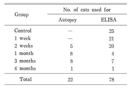
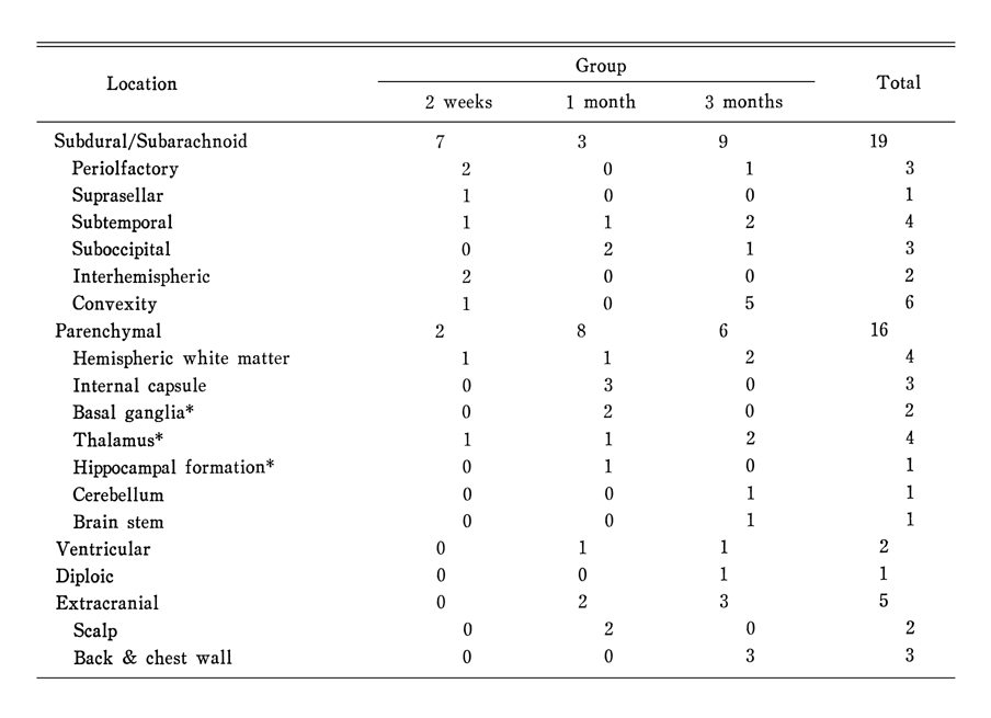
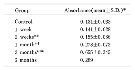
To establish an animal model of intracranial sparganosis, the fate and behavior of the experimentally inoculated spargana were observed. A total of 102 scolices of spargana were injected into 22 cat brains, and the cats were sacrificed at 2 weeks, 1 month, 3 months and 6 months after the inoculation. Neurosparganosis was established in 77% of the cats. Of 43 recovered worms, 19 (44%) were located in the subdural or subarachnoid space, 16 (37%) in the brain parenchyme, and 2 (5%) in the lateral ventricle. One was detected at the diploic space of the skull and 5 were outside the cranial cavity. All but one were alive, and had grown tails. They were distributed in the brain parenchyme randomly. There was no place which they could not invade. No adult was found in the intestine. Cerebrospinal fluid (CSF) was collected before inoculation, 1 week, 2 weeks, 1 month, 3 months and 6 months after inoculation. The level of anti-sparganum IgG antibody in CSF measured by ELISA began to increase above the criteria of positivity 1 month after inoculation. Three months after inoculation, the values markedly increased.
The present findings reveal that intracranial inoculation of spargana into the brains of cats would be a good animal model of experimental neurosparganosis.
Fig. 2 Gross finding of the brain and recovered worms. A; (3 months after inoculation) a worm on the suprasylvian gyrus (arrows), B; (2 weeks after inoculation) a worm in the interhemispheric fissure (arrow), C; (3 months after inoculation) a worm beneath the temporal lobe (arrow), D; (3 months after inoculation) growing worms with tail from the subdural space, E; (2 weeks after inoculation) a worm in the thalamus and worm tracts anterior and posterior to the worm, F; (1 months after inoculation) worms in the internal capsules of both sides and tracts including worms in the brain stem and the cerebellum, G; (3 months after inoculation) a worm recovered from the scalp (arrow), H; (3 months after inoculation) an worm recovered from the diploic space of the skull (small arrows) 5mm apart from the hole made for the inoculation of the worms (large arrow).
Fig. 3 Schematic drawing of the distribution of total recovered worms. Upper; worms in the subdural/subarachnoid (IH=interhemispheric fissure, AS=anterior sigmoid gyrus, L=lateral gyrus, SSV=suprasylvian gyrus, ES=ectosylvian gyrus, OB=olfactory bulb, SS=suprasellar cistern, ST=subtemporal, SO=suboccipital), Lower; intraparenchymal worms (WM=hemispheric white matter, LV=lateral ventricle, CN=caudate nucleus, IC=internal capsule, TH=thalamus, H=hippocampal formation, BS=brain stem, CBL=cerebellum)
Fig. 4 Results of ELISA on the cerebrospinal fluid (CSF, left) and the serum (right)
Anders K, Foley K, Stern E, Brown WJ. Intracranial sparganosis: an uncommon infection. Case report. J Neurosurg 1984;60(6):1282–1286.
2.
Anegawa S, Hayashi T, Ozuru K, Kuramoto S, Nishimura K, Shimizu T, Hirata M. Sparganosis of the brain. Case report. J Neurosurg 1989;71(2):287–289.
3.
Chang KH, Cho SY, Chi JG, Kim WS, Han MC, Kim CW, Myung H, Choi KS. Cerebral sparganosis: CT characteristics. Radiology 1987;165(2):505–510.
4.
Chi JG, Chi HS, Lee SH. Histopathologic Study On Human Sparganosis. Korean J Parasitol 1980;18(1):15–23.
5.
Cho SY, Bae JH, Seo BS. Some Aspects Of Human Sparganosis In Korea. Korean J Parasitol 1975;13(1):60–77.
6.
Cho SY, Kim SI, Kang SY, Park AJ. Intracranial synthesis of specific IgG antibody in cerebrospinal fluid of neurocysticercosis patients. Korean J Parasitol 1988;26(1):15–26.
7.
Cho SY, et al. Chung-Ang J Med 1982;7:321–333.
8.
Choi WJ. [Migration And Distribution Of Spargana In Body Of Experimentally Infected Mice]. Korean J Parasitol 1984;22(2):229–237.
9.
Choi WY, Yoo JE, Nam HW, Choi HR. [Purification of antigenic proteins of Paragonimus westermani and their applicability to experimental cat paragonimiasis]. Korean J Parasitol 1986;24(2):177–186.
10.
Corkum KC. Proc La Acad Sci 1973;36:64–70.
11.
Fan KJ, Pezeshkpour GH. Cerebral sparganosis. Neurology 1986;36(9):1249–1251.
12.
Han CH, et al. Chung-Ang J Med 1988;13(2):237–248.
13.
Hong ST, Kim KJ, Huh S, Lee YS, Chai JY, Lee SH, Lee YS. The changes of histopathology and serum anti-sparganum IgG in experimental sparganosis of mice. Korean J Parasitol 1989;27(4):261–269.
14.
Hong SB, et al. J Korean Neurol Ass 1985;3:96–101.
15.
Kim CH. [The Infection Status Of Sparganum And Gnathostoma In Frogs Of Southern Part Of Korea]. Korean J Parasitol 1983;21(1):83–86.
16.
Kim CH, Shin DW. [Prevalence of sparganum of frogs (Rana nigromaculata) in Dae-jeon area, Chung-nam, Korea]. Korean J Parasitol 1975;13(2):159–162.
17.
Kim H, Kim SI, Cho SY. Serological Diagnosis Of Human Sparganosis By Means Of Micro-ELISA. Korean J Parasitol 1984;22(2):222–228.
18.
Kim SC, et al. J Korean Neurosurg Soc 1981;10:589–593.
19.
Lee HB, et al. J Korean Neurol Ass 1987;5:64–69.
20.
Lee SH, Chai JY, Seo BS, Cho SY. Two cases of human infection by adult of Spirometra erinacei. Korean J Parasitol 1984;22(1):66–71.
21.
McLaren M, Draper CC, Roberts JM, Minter-Goedbloed E, Ligthart GS, Teesdale CH, Amin MA, Omer AH, Bartlett A, Voller A. Studies on the enzyme linked immunosorbent assay (ELISA) test for Schistosoma mansoni infections. Ann Trop Med Parasitol 1978;72(3):243–253.
22.
Mineura K, Mori T. Sparganosis of the brain. Case report. J Neurosurg 1980;52(4):588–590.
23.
Mueller JF. The biology of Spirometra. J Parasitol 1974;60(1):3–14.
24.
Youm JY, et al. J Korean Neurosurg Soc 1987;16:847–851.