Warning: mkdir(): Permission denied in /home/virtual/lib/view_data.php on line 81
Warning: fopen(upload/ip_log/ip_log_2024-05.txt): failed to open stream: No such file or directory in /home/virtual/lib/view_data.php on line 83
Warning: fwrite() expects parameter 1 to be resource, boolean given in /home/virtual/lib/view_data.php on line 84 Cercarial shedding of Echinostoma cinetorchis and experimental infection of the cercariae to several kinds of snails
Cercarial shedding of Echinostoma cinetorchis and experimental infection of the cercariae to several kinds of snails
Y K Ahn,Y S Ryang,*J Y Chai,** and W M Sohn***
*Department of Parasitology, Wonju College of Medicine and Department of Medical Technology, College of Health Sciences, Yonsei University, Wonju 220-050, Korea.
**Department of Parasitology, College of Medicine, Seoul National University, Seoul 110-460, Korea.
***Department of Parasitology, College of Medicine, Inje University, Pusan 614-112, Korea.
Abstract
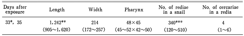
The development of Echinostoma cinetorchis in several snail species reared in laboratory aquaria was observed. The eggs from adult flukes collected from the intestine of rats were cultivated to miracidia, and exposed to Hippeutis sp. snails. Observations were made for cercarial shedding from the exposed snails. The cercariae shed from the snails were again exposed to several species of fresh water snails in order to observe metacercarial formation in the snails and their infectivity to final hosts. The results obtained in this study were as follows: 1. Twenty miracidia were exposed to each snail of Hippeutis sp. About 58.3% of the above snails (7 out of 12) were dead before shedding the cercariae, and the remainder shed the cercariae for a period of 7 to 9 days before death. 2. Cercarial shedding from the infected snails started from the 25th day after the exposure to miracidia, and the total number of cercariae shed per snail was 684 in average (range; 482-904). 3. The size of rediae developed in the infected Hippeutis sp. snails was 1,242 × 214 µm in average, and the number of rediae per snail was 350 in average (range; 120-510). 4. About 40 to 50 cercariae shed from the Hippeutis sp. snails were each exposed to several species of snails reared in the laboratory. The metacercarial formation was confirmed by dissecting the infected snails, 12 to 16 days after the infection.
Figures
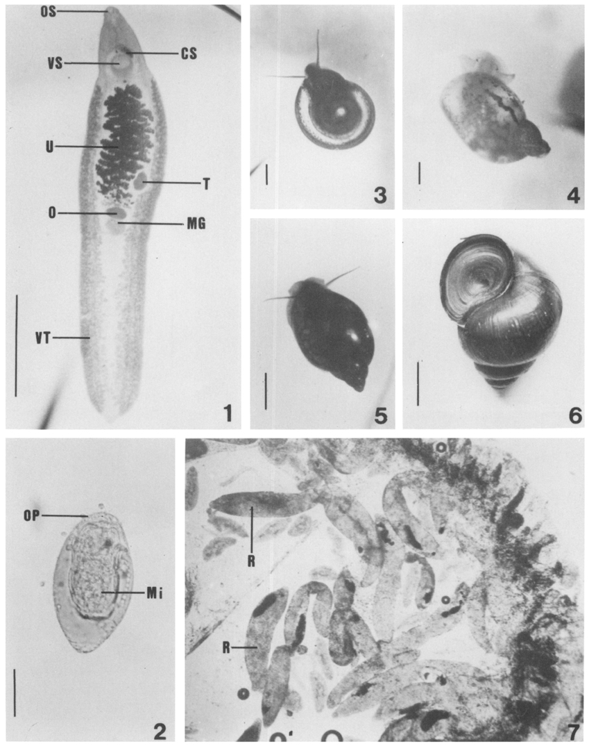
Figs. 1-7 Fig. 1. Adult E. cinetorchis recovered 30 days after infection, showing oral (OS) and ventral suckers (VS), cirrus sac (CS), uterus (U) with numerous eggs, Mehlis' gland (MG), ovary (O), vitellaria (VT) and a moved testis (T). (scale : 3mm)
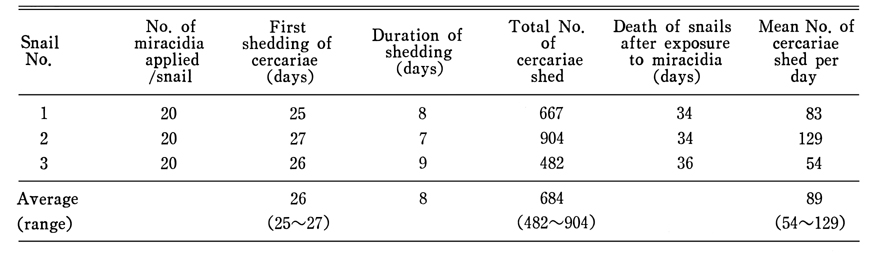
Fig. 2. A mature egg, with fully developed miracidium (Mi) and operculum (P) 3 weeks after cultivation. (scale : 30 µm)
Fig. 3~5. Adult snails of Hippeutis sp. (fig. 3), Radix auricularia coreana (Fig. 4) and Physa acuta (Fig. 5), reared for 2.5 months in our laboratory. (scale : 3mm)
Fig. 6. Mud-snail, Cipangopaludina sp. collected from the field. (scale : 10mm)
Fig. 7. Rediae (R) containing growing cercariae in the snail (first intermediate host), Hippeutis sp., 20~25 day post-exposure to miracidia.
Figs. 8-10 Fig. 8. A mature redia, containing motile cercariae in the body cavity (C: cercaria, IC: intestinal cecum, LP: lateral prosess, UC: undeveloped cercaria). (scale : 200 µm)
Fig. 9. Vigorous cercariae under a cover slip, discharged immediately from redia. (a: constricted shape, b : extended shape. COD: circumoral disc, EG: mass of excretory granules)
Fig. 10.E. cinetorchis metacercariae (Me) encysted in snails, Radix sp. (a) and Cipagopaludina sp. (b). (scale : 100 µm)
Tables
Table 1 Cercarial shedding of Hippeutis sp. snails exposed to the E. cinetorchis miracidia (water temp. : 26~28℃)
Table 2 Daily cercarial shedding of E. cinetorchis from experimentally infected snails (Hippeutis sp.) (water temp. : 26~28℃)
Table 3 Shedding pattern of E. cinetorchis cercariae from experimentally infected snails (Hippeutis sp.) (water temp. : 26~28℃)
Table 4 Measurenents of rediae in snails (Hippeutis sp.) experimentally infected with the miracidia of E. cinetorchis (unite : µm) (water temp. : 26~28℃)
Table 5 Infectivity and density of metacercariae encysted in various kinds of snails experimentally infected with the cercariae of E. cinetorchis
References
2.
Ando R. Nisshin Igaku 1938;27(12):1717–1737.
3.
Ando R, et al. Dobutsugaku Zasshi 1923;35:108–119.
5.
Bae KH. J Kurume Med Assoc 1983;46(9):793–818.
6.
Burch JB, et al. Walkerana 1987;2(8):195–232.
7.
Cho SY, Kang SY, Ryang YS. [Helminthes Infections In The Small Intestine Of Stray Dogs In Ejungbu City, Kyunggi Do, Korea]. Korean J Parasitol 1981;19(1):55–59.
9.
Koga M. Nippon Kiseichu Gakkai Kiji 1938;10:83.
10.
Kwon OK, et al. Korean J Limnol 1982;5(12):39–50.
11.
Kurisu Y. Kummamoto Igakkai Zasshi 1932;8(4):283–298.
13.
Lee SK, Chung NS, Ko IH, Ko HI, Sohn WM. [A case of natural human infection by Echinostoma cinetorchis]. Korean J Parasitol 1988;26(1):61–64.
14.
Miki T. Aichi Igakkai Zasshi 1923;30(3):499–504.
16.
Ryang YS, Ahn KY, Kim WT, Shin KC, Lee KW, Kim TS. [Two cases of human infection by Echinostoma cinetorchis]. Korean J Parasitol 1986;24(1):71–76.
17.
Seo BS, et al. Seoul J Med 1980;21(1):21–29.
18.
Seo BS, Cho SY, Hong ST, Hong SJ, Lee SH. Studies On Parasitic Helminths Of Korea 5.Survey On Intestinal Trematodes Of House Rats. Korean J Parasitol 1981;19(2):131–136.
19.
Seo BS, Park YH, Chai JY, Hong SJ, Lee SH. [Studies On Intestinal Trematodes In Korea Xiv. Infection Status Of Loaches With Metacercariae Of Echinostoma Cinetorchis And Their Development In Albino Rats]. Korean J Parasitol 1984;22(2):181–189.
20.
Seo BS, Rim HJ, Lee CW. Studies on the parasitic helmiths of Korea: I. Trematodes of rodents. Korean J Parasitol 1964;2(1):20–26.
21.
Takahashi S. Fukuoka Ika Daigaku Zasshi 1927;20(6):712–723.
22.
Yamashita J. Progress of Med Parasit in Jpn 1964;1:288–313.